

One of the biggest challenges in psychiatric and neurological research is figuring out what is happening in the living human brain at the molecular level. Traditionally, most studies have relied on postmortem brain tissue because collecting brain samples from living people is not possible in most cases. But is gene expression in postmortem tissue really the same as in a living brain?
Researchers from the Icahn School of Medicine at Mount Sinai tackled this important question using RNA sequencing. They compared gene expression in the prefrontal cortex between 275 living samples and 243 postmortem samples. The results were striking. Nearly 80 percent of genes showed significant differences between the two groups.
The Living Brain Project (LBP)
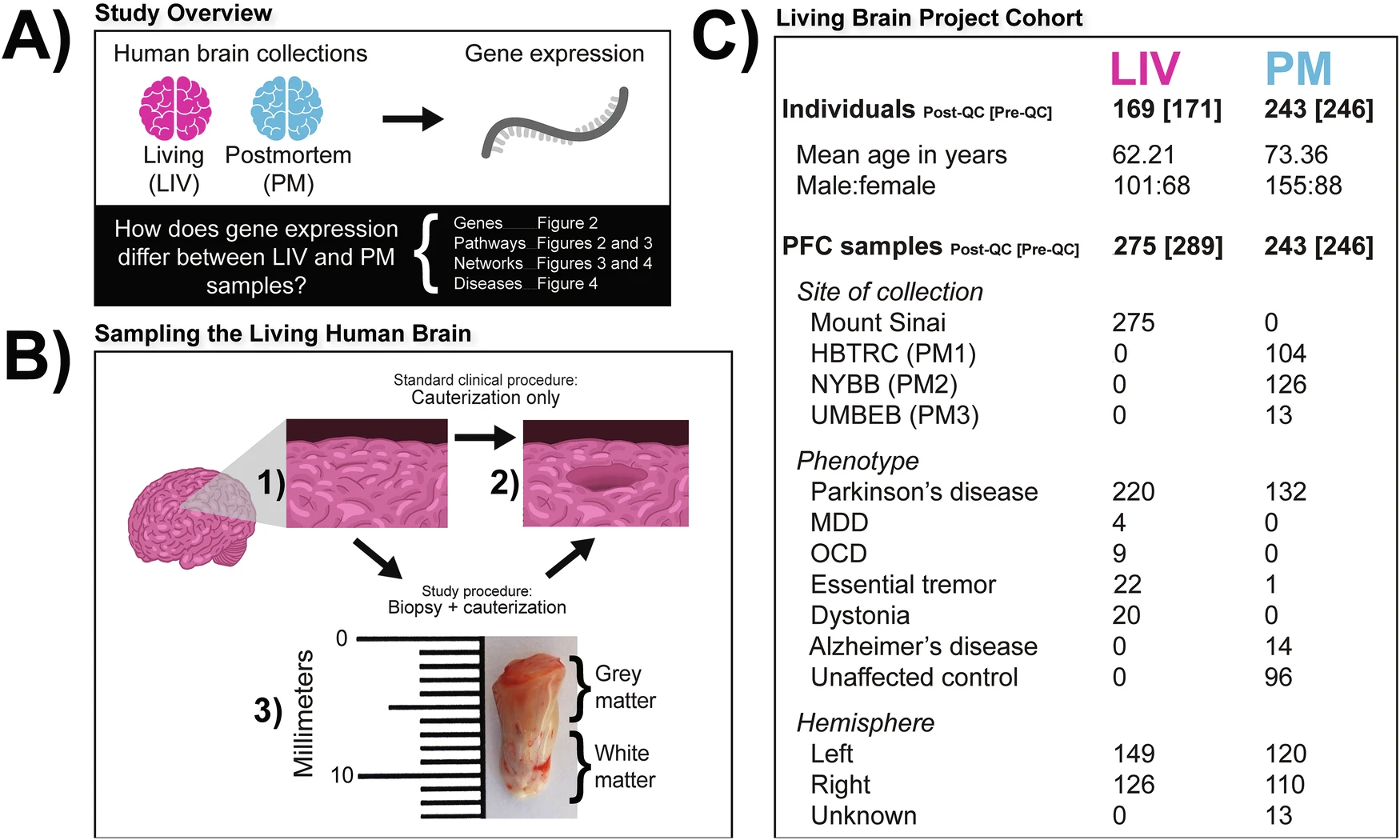
A Study overview. The pink and blue colors represent LIV samples and PM samples, respectively, throughout all subsequent figures. B Sampling the living human brain. The schematic illustrates that the standard clinical procedure and the modified study procedure amount to the same effective PFC volume loss. In (1) the state of the PFC at baseline is depicted. In (2) the final state of the PFC is depicted (i.e., the inner cylinder of tissue removed). The arrows and associated labels indicate how the final state of preparation is achieved in the standard clinical procedure (i.e., cauterization only) and the LBP procedure (i.e., biopsy followed by cauterization). Shown in (3) is a photograph of a PFC biopsy obtained for the LBP that is representative of the samples studied in this report. The top of the image is the anterior surface of the PFC. C Living Brain Project cohort. Numbers refer to sample size (i.e., individuals or samples) except for age. The individual and sample numbers are identical for the PM cohort because only one sample was obtained per individual. Sample sizes inside and outside of the square brackets indicate counts prior to and after the application of quality control filters, respectively. [PFC prefrontal cortex, QC quality control, HBTRC (PM1) Harvard Brain and Tissue Resource Center, NYBB (PM2) New York Brain Bank at Columbia University, UMBEB (PM3) University of Miami Brain Endowment Bank, MDD major depressive disorder, OCD obsessive-compulsive disorder].
The researchers worked carefully to rule out other explanations, such as differences in cell type, RNA quality, postmortem delay, age, medications, or even the computational tools used. None of these factors explained the differences. To confirm their findings, they analyzed data from two additional independent cohorts, showing that the gene expression differences were real and consistent across all brain cell types.
This discovery has major implications. It suggests that gene expression signatures seen in postmortem studies, whether related to psychiatric disorders, neurological conditions, or even normal traits like aging, may not always reflect what is actually happening in the living brain. While postmortem tissue will remain useful for many types of research, future studies using safely collected living brain tissue could open new opportunities to study real-time processes, such as how the brain responds to emotions at the molecular level.
By highlighting the differences between living and postmortem gene expression, this research provides a roadmap for how future psychiatric and neurological research can more accurately study human brain health and illness.
Availability – All data analyzed from the full LBP cohort are available via the AD Knowledge Portal (https://adknowledgeportal.org).
Liharska L, Park Y, Ziafat K, Wilkins L, Silk H, Linares L, Thompson R, Vornholt E, Sullivan B, Cohen V, Kota P, Feng C, Cheng E, Johnson J, Rieder M, Huang J, Scarpa J, Polanco J, Moya E, Hashemi A, Levin M, Nadkarni G, Sebra R, Crary J, Schadt E, Beckmann N, Kopell B, Charney A. (2025) A study of gene expression in the living human brain. Molecular Psychiatry [article]
Related Posts






One of the biggest challenges in psychiatric and neurological research is figuring out what is happening in the living human brain at the molecular level. Traditionally, most studies have relied on postmortem brain tissue because collecting brain samples from living people is not possible in most cases. But is gene expression in postmortem tissue really the same as in a living brain?
Researchers from the Icahn School of Medicine at Mount Sinai tackled this important question using RNA sequencing. They compared gene expression in the prefrontal cortex between 275 living samples and 243 postmortem samples. The results were striking. Nearly 80 percent of genes showed significant differences between the two groups.
The Living Brain Project (LBP)
A Study overview. The pink and blue colors represent LIV samples and PM samples, respectively, throughout all subsequent figures. B Sampling the living human brain. The schematic illustrates that the standard clinical procedure and the modified study procedure amount to the same effective PFC volume loss. In (1) the state of the PFC at baseline is depicted. In (2) the final state of the PFC is depicted (i.e., the inner cylinder of tissue removed). The arrows and associated labels indicate how the final state of preparation is achieved in the standard clinical procedure (i.e., cauterization only) and the LBP procedure (i.e., biopsy followed by cauterization). Shown in (3) is a photograph of a PFC biopsy obtained for the LBP that is representative of the samples studied in this report. The top of the image is the anterior surface of the PFC. C Living Brain Project cohort. Numbers refer to sample size (i.e., individuals or samples) except for age. The individual and sample numbers are identical for the PM cohort because only one sample was obtained per individual. Sample sizes inside and outside of the square brackets indicate counts prior to and after the application of quality control filters, respectively. [PFC prefrontal cortex, QC quality control, HBTRC (PM1) Harvard Brain and Tissue Resource Center, NYBB (PM2) New York Brain Bank at Columbia University, UMBEB (PM3) University of Miami Brain Endowment Bank, MDD major depressive disorder, OCD obsessive-compulsive disorder].
The researchers worked carefully to rule out other explanations, such as differences in cell type, RNA quality, postmortem delay, age, medications, or even the computational tools used. None of these factors explained the differences. To confirm their findings, they analyzed data from two additional independent cohorts, showing that the gene expression differences were real and consistent across all brain cell types.
This discovery has major implications. It suggests that gene expression signatures seen in postmortem studies, whether related to psychiatric disorders, neurological conditions, or even normal traits like aging, may not always reflect what is actually happening in the living brain. While postmortem tissue will remain useful for many types of research, future studies using safely collected living brain tissue could open new opportunities to study real-time processes, such as how the brain responds to emotions at the molecular level.
By highlighting the differences between living and postmortem gene expression, this research provides a roadmap for how future psychiatric and neurological research can more accurately study human brain health and illness.
Availability – All data analyzed from the full LBP cohort are available via the AD Knowledge Portal (https://adknowledgeportal.org).
Liharska L, Park Y, Ziafat K, Wilkins L, Silk H, Linares L, Thompson R, Vornholt E, Sullivan B, Cohen V, Kota P, Feng C, Cheng E, Johnson J, Rieder M, Huang J, Scarpa J, Polanco J, Moya E, Hashemi A, Levin M, Nadkarni G, Sebra R, Crary J, Schadt E, Beckmann N, Kopell B, Charney A. (2025) A study of gene expression in the living human brain. Molecular Psychiatry [article]
Related Posts
Recent Posts
 RNAmel simplifies RNA sequencing data analysis with a reproducible, integrated platform
 Heart failure reshapes bone marrow to create inflammation-related immune memory
 Overlooked DNA regions surrounding ‘microexons’ hold missing pieces of autism’s genetic puzzle
 RiboBA improves detection of hidden protein-coding regions using ribosome profiling
 An anti-senescence gene may fight spinal disc deterioration
 Bulk RNA-seq Analysis: A Complete Beginner’s Guide
RNA-Seq Products & Services


Stay Connected