

T cell immunotherapy, a cutting-edge approach in fighting cancer and infectious diseases, relies on finding the right T cell receptors (TCRs) to target harmful antigens. TCRs are like “keys” that fit into specific “locks” on harmful cells, activating T cells to attack and destroy them. Traditionally, scientists have used a method called MHC multimer staining to identify these antigen-reactive TCRs. However, this method often misses TCRs that may not bind strongly to antigens but are still highly effective in activating T cells for the fight.
To address this limitation, researchers at the Children’s Hospital of Philadelphia have developed a groundbreaking technology called ATLAS-seq (Aptamer-based T Lymphocyte Activity Screening and SEQuencing). This method uses a tiny, high-tech device called a microfluidic platform to identify T cells that react strongly to specific antigens. What makes ATLAS-seq unique is its use of a fluorescent sensor that detects the release of a molecule called IFNγ, which is a clear signal that a T cell is actively responding to an antigen.
Here’s how it works:
- Single-Cell Precision: The microfluidic platform isolates individual T cells and exposes them to antigens.
- Detecting Activity: If a T cell reacts, the sensor lights up, showing that it’s secreting IFNγ.
- Deep Analysis: Researchers then perform single-cell RNA sequencing and TCR sequencing to understand the T cell’s genetic and functional makeup.
Aptamer-based T-lymphocyte activity screening and sequencing (ATLAS-seq)
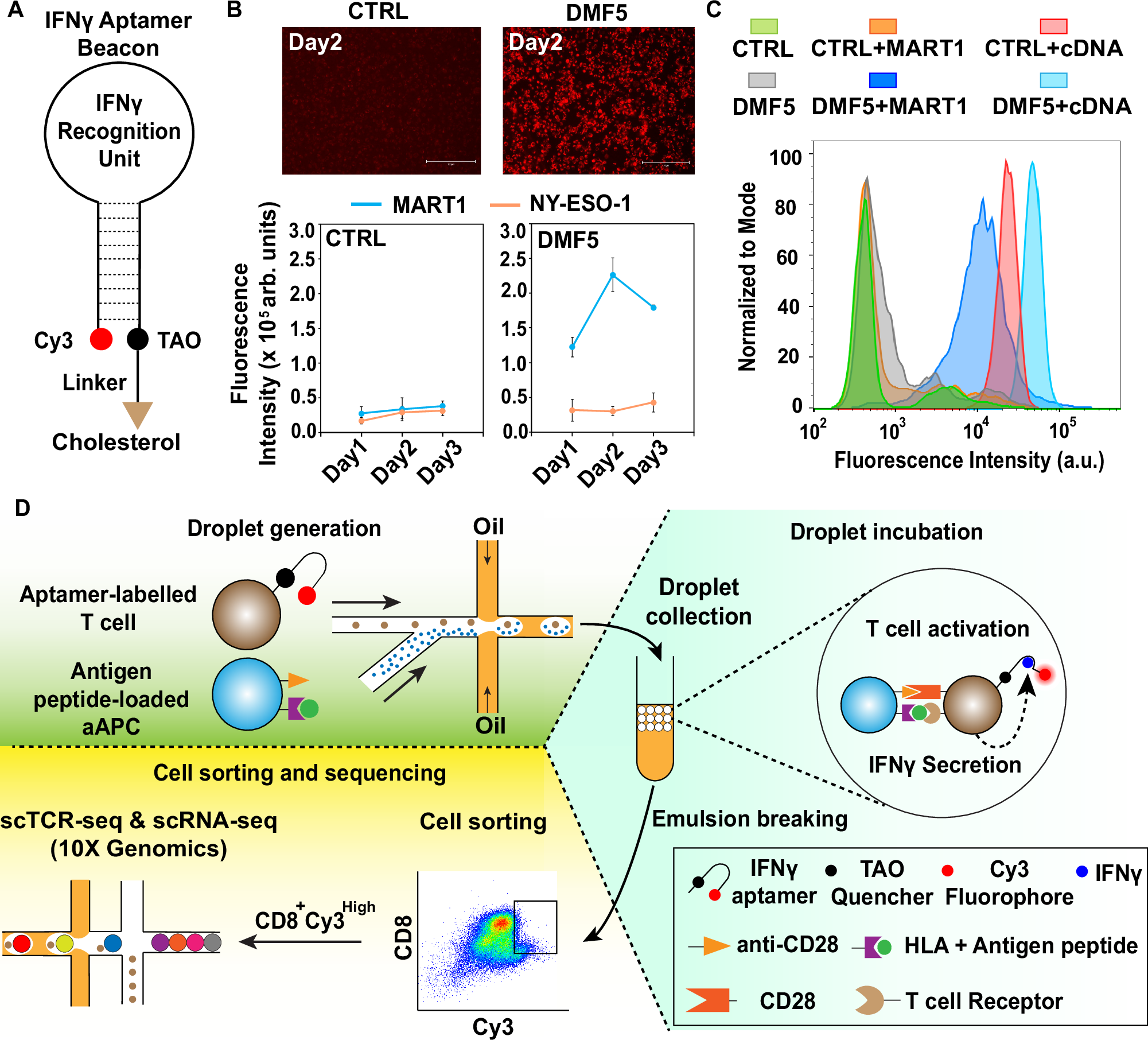
A Stem-loop structure of IFNγ-specific aptamer beacon. The 5’ end of the aptamer is decorated with the Cy3 fluorophore. The 3’ end of the aptamer is decorated with the TAO quencher, iSP18 linker and cholesterol. B MART1 or NY-ESO-1 peptide-loaded aAPCs were added to 5 × 10⁵ cells/well DMF5 Jurkat cells or control (CTRL) Jurkat cells decorated with the IFNγ-specific aptamer beacons (1nmol/106 cells) at 2:1 ratio in 96-well plate. Top: Representative images of control or DMF5 Jurkat cells treated with MART1 aAPCs on day 2. Bottom: Average cellular fluorescence intensities of the aptamer beacons were measured to detect IFNγ secretion due to MART1 antigen-specific DMF5 Jurkat T cell activation. mean ± s.d. of n = 3 independent biological replicates. Arb. Units: arbitrary units. C Control (CTRL) Jurkat and DMF5 Jurkat cells were decorated with IFNγ-specific aptamer beacons (1nmol/106 cells) and co-encapsulated in droplets with MART1 peptide-loaded aAPCs or empty aAPCs for 2 days at 37 °C, 5% CO2. Fluorescence intensities of aptamer beacons were measured by flow cytometry to analyze the IFNγ secretion from MART1 antigen-specific activated single Jurkat cells in droplets. Aptamer beacon-decorated control Jurkat cells: with empty aAPCs (green); with MART1 peptide-loaded aAPCs (orange); with aptamer cDNA (red). Aptamer beacon-decorated DMF5 Jurkat cells: with empty aAPCs (gray); with MART1 peptide-loaded aAPCs (dark blue); with aptamer cDNA (light blue). Aptamer cDNA: DNA oligo with a complementary sequence to the aptamer stem region. D ATLAS-seq workflow. A single aptamer-labeled T cell is co-encapsulated with antigen peptide-loaded aAPC beads within uniformly-sized water-in-oil microdroplets using a cross junction channel in a microfluidic chip. Droplets are incubated off-chip to accumulate IFNγ secreted from the encapsulated single T cell. Secreted IFNγ will switch on the Cy3 fluorescence signal of the aptamer beacon on the T cell surface. After breaking the emulsion, recovered T cells are analyzed and sorted by flow cytometry based on the intensity of Cy3 fluorescence signal. CD8+/Cy3high T cells are used for preparing both single-cell TCR-seq and single-cell RNA-seq libraries using 10X Genomics Chromium Controller.
In a recent study, the scientists used ATLAS-seq to identify TCRs reactive to cytomegalovirus (CMV) and prostate-specific antigen (PSA). They discovered that TCRs identified through ATLAS-seq were better at activating T cells and killing target cells compared to those found using traditional MHC multimer staining. This means ATLAS-seq can uncover highly effective TCRs that other methods might miss.
The implications of this technology are enormous. By providing a more efficient and accurate way to screen for antigen-reactive TCRs, ATLAS-seq could accelerate the development of engineered T cell therapies. This is especially crucial for conditions like cancer, where finding the right TCRs can make a significant difference in treatment outcomes.
In short, ATLAS-seq represents a powerful new tool in the fight against disease, offering hope for more effective and personalized immunotherapies.
Luo S, Notaro A, Lin L. (2025) ATLAS-seq: a microfluidic single-cell TCR screen for antigen-reactive TCRs. Nat Commun 16(1):216. [article]
Related Posts






T cell immunotherapy, a cutting-edge approach in fighting cancer and infectious diseases, relies on finding the right T cell receptors (TCRs) to target harmful antigens. TCRs are like “keys” that fit into specific “locks” on harmful cells, activating T cells to attack and destroy them. Traditionally, scientists have used a method called MHC multimer staining to identify these antigen-reactive TCRs. However, this method often misses TCRs that may not bind strongly to antigens but are still highly effective in activating T cells for the fight.
To address this limitation, researchers at the Children’s Hospital of Philadelphia have developed a groundbreaking technology called ATLAS-seq (Aptamer-based T Lymphocyte Activity Screening and SEQuencing). This method uses a tiny, high-tech device called a microfluidic platform to identify T cells that react strongly to specific antigens. What makes ATLAS-seq unique is its use of a fluorescent sensor that detects the release of a molecule called IFNγ, which is a clear signal that a T cell is actively responding to an antigen.
Here’s how it works:
- Single-Cell Precision: The microfluidic platform isolates individual T cells and exposes them to antigens.
- Detecting Activity: If a T cell reacts, the sensor lights up, showing that it’s secreting IFNγ.
- Deep Analysis: Researchers then perform single-cell RNA sequencing and TCR sequencing to understand the T cell’s genetic and functional makeup.
Aptamer-based T-lymphocyte activity screening and sequencing (ATLAS-seq)
A Stem-loop structure of IFNγ-specific aptamer beacon. The 5’ end of the aptamer is decorated with the Cy3 fluorophore. The 3’ end of the aptamer is decorated with the TAO quencher, iSP18 linker and cholesterol. B MART1 or NY-ESO-1 peptide-loaded aAPCs were added to 5 × 10⁵ cells/well DMF5 Jurkat cells or control (CTRL) Jurkat cells decorated with the IFNγ-specific aptamer beacons (1nmol/106 cells) at 2:1 ratio in 96-well plate. Top: Representative images of control or DMF5 Jurkat cells treated with MART1 aAPCs on day 2. Bottom: Average cellular fluorescence intensities of the aptamer beacons were measured to detect IFNγ secretion due to MART1 antigen-specific DMF5 Jurkat T cell activation. mean ± s.d. of n = 3 independent biological replicates. Arb. Units: arbitrary units. C Control (CTRL) Jurkat and DMF5 Jurkat cells were decorated with IFNγ-specific aptamer beacons (1nmol/106 cells) and co-encapsulated in droplets with MART1 peptide-loaded aAPCs or empty aAPCs for 2 days at 37 °C, 5% CO2. Fluorescence intensities of aptamer beacons were measured by flow cytometry to analyze the IFNγ secretion from MART1 antigen-specific activated single Jurkat cells in droplets. Aptamer beacon-decorated control Jurkat cells: with empty aAPCs (green); with MART1 peptide-loaded aAPCs (orange); with aptamer cDNA (red). Aptamer beacon-decorated DMF5 Jurkat cells: with empty aAPCs (gray); with MART1 peptide-loaded aAPCs (dark blue); with aptamer cDNA (light blue). Aptamer cDNA: DNA oligo with a complementary sequence to the aptamer stem region. D ATLAS-seq workflow. A single aptamer-labeled T cell is co-encapsulated with antigen peptide-loaded aAPC beads within uniformly-sized water-in-oil microdroplets using a cross junction channel in a microfluidic chip. Droplets are incubated off-chip to accumulate IFNγ secreted from the encapsulated single T cell. Secreted IFNγ will switch on the Cy3 fluorescence signal of the aptamer beacon on the T cell surface. After breaking the emulsion, recovered T cells are analyzed and sorted by flow cytometry based on the intensity of Cy3 fluorescence signal. CD8+/Cy3high T cells are used for preparing both single-cell TCR-seq and single-cell RNA-seq libraries using 10X Genomics Chromium Controller.
In a recent study, the scientists used ATLAS-seq to identify TCRs reactive to cytomegalovirus (CMV) and prostate-specific antigen (PSA). They discovered that TCRs identified through ATLAS-seq were better at activating T cells and killing target cells compared to those found using traditional MHC multimer staining. This means ATLAS-seq can uncover highly effective TCRs that other methods might miss.
The implications of this technology are enormous. By providing a more efficient and accurate way to screen for antigen-reactive TCRs, ATLAS-seq could accelerate the development of engineered T cell therapies. This is especially crucial for conditions like cancer, where finding the right TCRs can make a significant difference in treatment outcomes.
In short, ATLAS-seq represents a powerful new tool in the fight against disease, offering hope for more effective and personalized immunotherapies.
Luo S, Notaro A, Lin L. (2025) ATLAS-seq: a microfluidic single-cell TCR screen for antigen-reactive TCRs. Nat Commun 16(1):216. [article]
Related Posts
Recent Posts
 Machine learning reveals how RNA splicing is regulated in individual cell types
 Illumina releases SpliceAI2 to help advance rare disease research
 Postdoctoral Fellow – Long-Read RNA Methods Development for Neurodegeneration
 Novel biosensor uses DNA ‘barcodes’ to uncover disease signals in cells
 GP-DHT uses single-cell RNA sequencing to map gene regulatory networks
 Boehringer taps Envisagenics’ AI to mine RNA splicing for cancer targets
RNA-Seq Products & Services



Stay Connected