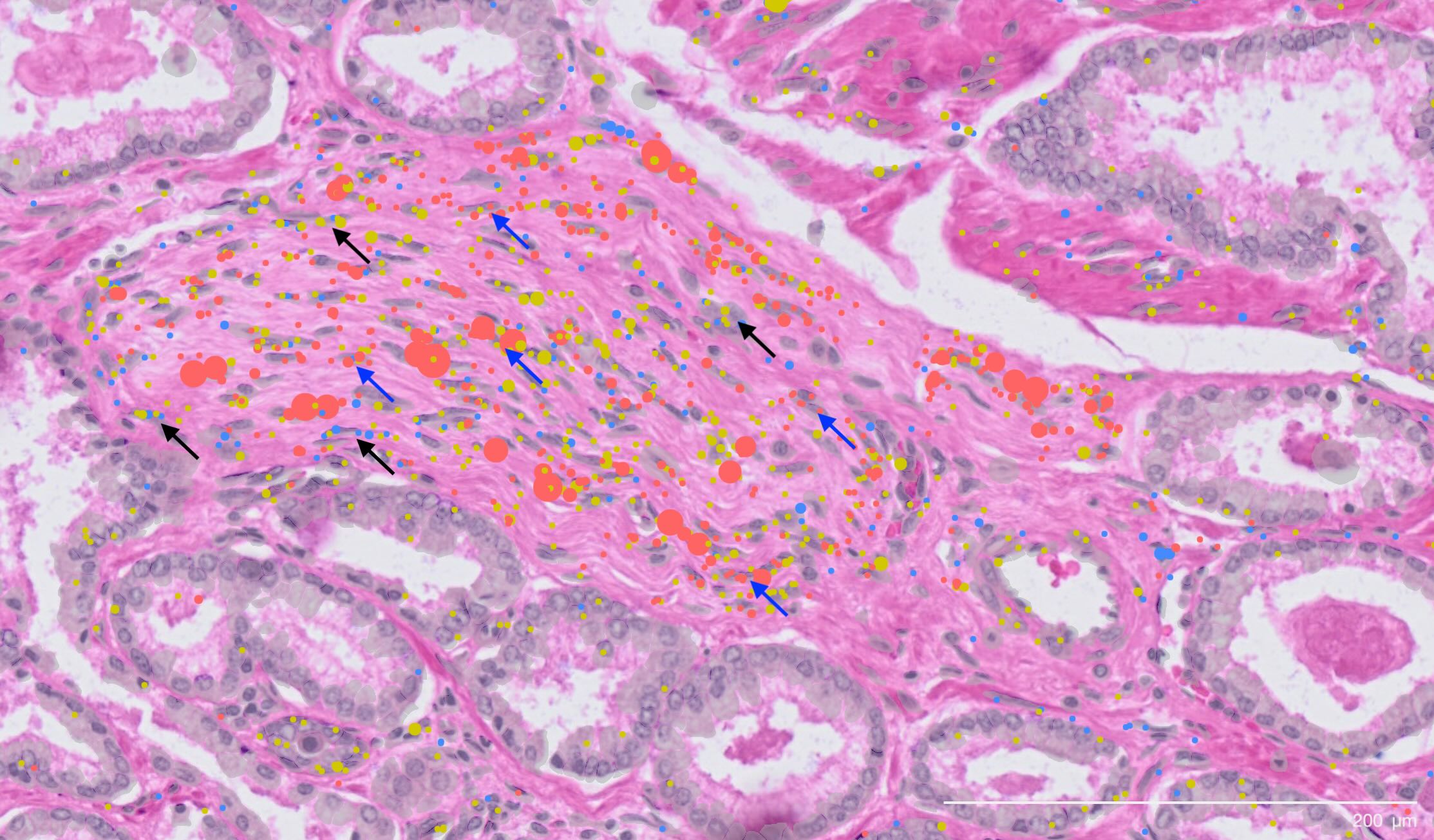
The newly discovered cell type (yellow) shown clustering around a prostate cancer nerve bundle.
Prostate cancer affects one in five Australian men, making it the most common cancer in the country. Now, researchers at the Garvan Institute of Medical Research have produced the world’s most detailed cellular ‘atlas’ of early-stage prostate cancer, revealing the earliest changes that lead to the disease.
The study describes a never-before-identified cell type and shows that many cells appearing healthy under the microscope have already begun their journey towards malignancy. These findings could reveal new risk factors, lead to earlier detection methods and help determine which patients are at risk of developing aggressive disease. The study was published in Cancer Research, a journal of the American Association for Cancer Research.
The research team, led by Professor Alexander Swarbrick, Co-Director of the Cancer Ecosystems Program and Lab Head at Garvan, analysed tissue from 24 patients recently diagnosed with early-stage prostate cancer. The samples were drawn from the Garvan St Vincent’s Prostate Cancer Biobank – the largest in the southern hemisphere, with contributions from more than 16,000 patients over three decades. The team then used advanced single-cell RNA sequencing to examine the genetic activity of every single cell in a tissue, and spatial transcriptomics to map their exact locations in the tumour landscape.
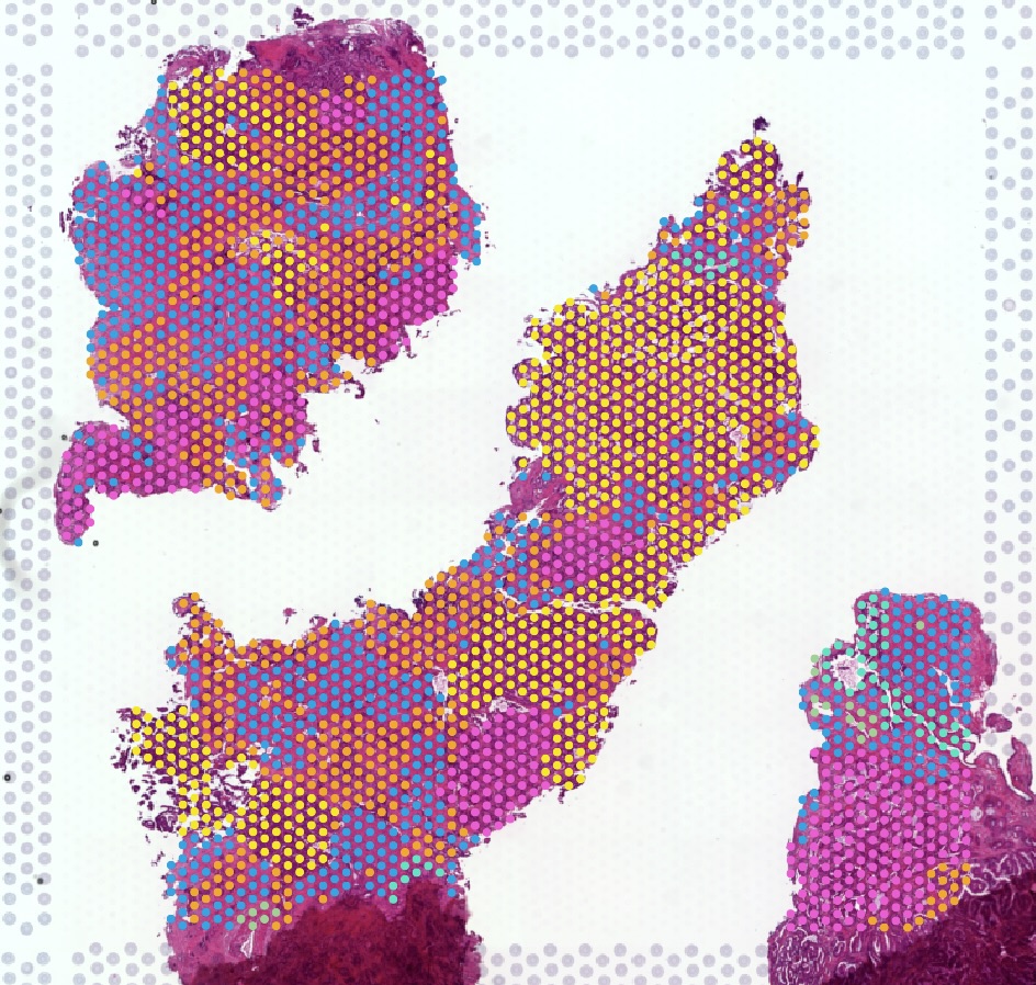
Spatial mapping reveals the complex cellular landscape of prostate tumours
This resulted in a comprehensive atlas, identifying 11 major cell types, 50 minor subtypes, and numerous cell state classifications within prostate cancer tissue. This level of detail provides the global research community with an invaluable resource for studying how the tumours form and opportunities to design new therapeutics.
“We’ve captured a molecular readout showing years of cancer development,” says Professor Swarbrick. “What’s particularly surprising is the significant number of cells that look perfectly ordinary but have already acquired DNA changes associated with cancer. This suggests that current diagnostic methods using conventional microscopy may not be granular enough to detect these early mutations.”
Implications for early detection
Standard diagnosis relies on visually identifying structural irregularities in cells under a microscope. However, this study revealed that the progression from healthy prostate tissue to cancer involves multiple cellular states that are invisible. The researchers found that many epithelial cells – the cells that line organs and are where most cancers originate – showed early genomic alterations, even in tissue that appeared ‘normal’. This suggests that molecular testing could one day be used to detect these invisible warning signs, potentially allowing for diagnosis before the cancer can be seen by a pathologist.
Clinical lead Professor Anthony Joshua, Lab Head at Garvan and Head of the Department of Medical Oncology at St Vincent’s Hospital Sydney, emphasises the clinical significance of these findings. “Currently we don’t know how prostate cancer starts. Understanding the sequence of events that lead to life-threatening disease – and finding the signatures of cancer in normal-looking cells – is essential for creating better predictive tests and therapies.
“While most prostate cancer patients respond well to current treatments, this atlas provides foundational knowledge of the disease’s first stages that could help us pinpoint those at high risk of dangerous progression, who need more intensive intervention,” he says.
Surprise discovery of new cell type
Among the study’s most significant findings is the identification of a previously unknown cell type – called perineural cancer-associated fibroblasts, a type of connective tissue cell located near nerves in cancers.
“These perineural fibroblasts in tumours appear to have specialised machinery to communicate with nerves. We don’t yet know what these cells are doing, but the role of nerves in tumours is getting increasing attention in the cancer field. We’ve been able to classify these cells at the molecular level for the first time, giving them a distinct identity,” says Professor Swarbrick.
The tendency for tumours to grow in and around nerves in prostate cancer has long been associated with worse patient outcomes. This discovery of specialised cells that interact with these nerves provides a new piece of the puzzle in understanding how prostate tumours may recruit nerves, or spread to other parts of the body along the nerves to cause metastatic disease.
Next steps
The research team plans to expand the number of patients studied and investigate specific cellular populations, particularly those cells that look healthy but harbour genetic changes.
“We’ve had a peek through the window, but it’s still somewhat blurry,” says Professor Swarbrick. “Now we have more precise questions about these pre-malignant cells, and we can apply more focused techniques to be crystal clear about the sequence of genetic events that drive this cancer.”
Future work will also focus on understanding the function of the newly discovered perineural fibroblasts, which may reveal why prostate tumours that contain more nerves have worse outcomes.
While the team emphasises this is fundamental science rather than of immediate clinical application, the findings pave the way for future development of predictive tests. Given the high incidence of prostate cancer, even small improvements in early detection and treatment selection could benefit thousands of Australians.
Source – Garvan Institute of Medical Research


The newly discovered cell type (yellow) shown clustering around a prostate cancer nerve bundle.
Prostate cancer affects one in five Australian men, making it the most common cancer in the country. Now, researchers at the Garvan Institute of Medical Research have produced the world’s most detailed cellular ‘atlas’ of early-stage prostate cancer, revealing the earliest changes that lead to the disease.
The study describes a never-before-identified cell type and shows that many cells appearing healthy under the microscope have already begun their journey towards malignancy. These findings could reveal new risk factors, lead to earlier detection methods and help determine which patients are at risk of developing aggressive disease. The study was published in Cancer Research, a journal of the American Association for Cancer Research.
The research team, led by Professor Alexander Swarbrick, Co-Director of the Cancer Ecosystems Program and Lab Head at Garvan, analysed tissue from 24 patients recently diagnosed with early-stage prostate cancer. The samples were drawn from the Garvan St Vincent’s Prostate Cancer Biobank – the largest in the southern hemisphere, with contributions from more than 16,000 patients over three decades. The team then used advanced single-cell RNA sequencing to examine the genetic activity of every single cell in a tissue, and spatial transcriptomics to map their exact locations in the tumour landscape.
Spatial mapping reveals the complex cellular landscape of prostate tumours
This resulted in a comprehensive atlas, identifying 11 major cell types, 50 minor subtypes, and numerous cell state classifications within prostate cancer tissue. This level of detail provides the global research community with an invaluable resource for studying how the tumours form and opportunities to design new therapeutics.
Implications for early detection
Standard diagnosis relies on visually identifying structural irregularities in cells under a microscope. However, this study revealed that the progression from healthy prostate tissue to cancer involves multiple cellular states that are invisible. The researchers found that many epithelial cells – the cells that line organs and are where most cancers originate – showed early genomic alterations, even in tissue that appeared ‘normal’. This suggests that molecular testing could one day be used to detect these invisible warning signs, potentially allowing for diagnosis before the cancer can be seen by a pathologist.
Clinical lead Professor Anthony Joshua, Lab Head at Garvan and Head of the Department of Medical Oncology at St Vincent’s Hospital Sydney, emphasises the clinical significance of these findings. “Currently we don’t know how prostate cancer starts. Understanding the sequence of events that lead to life-threatening disease – and finding the signatures of cancer in normal-looking cells – is essential for creating better predictive tests and therapies.
Surprise discovery of new cell type
Among the study’s most significant findings is the identification of a previously unknown cell type – called perineural cancer-associated fibroblasts, a type of connective tissue cell located near nerves in cancers.
The tendency for tumours to grow in and around nerves in prostate cancer has long been associated with worse patient outcomes. This discovery of specialised cells that interact with these nerves provides a new piece of the puzzle in understanding how prostate tumours may recruit nerves, or spread to other parts of the body along the nerves to cause metastatic disease.
Next steps
The research team plans to expand the number of patients studied and investigate specific cellular populations, particularly those cells that look healthy but harbour genetic changes.
Future work will also focus on understanding the function of the newly discovered perineural fibroblasts, which may reveal why prostate tumours that contain more nerves have worse outcomes.
While the team emphasises this is fundamental science rather than of immediate clinical application, the findings pave the way for future development of predictive tests. Given the high incidence of prostate cancer, even small improvements in early detection and treatment selection could benefit thousands of Australians.
Source – Garvan Institute of Medical Research
Apostolov E, Roden DL, Holliday H, Cazet A, Harvey K, Zhang H, Wu SZ, van der Leij S, Kim HJ, Selth LA, Bartonicek N, Al-Eryani G, Reeves JL, He M, Lundeberg J, Potter AJ, Kench JG, Stricker PD, Joshua AM, Horvath LG, Swarbrick A. (2026) Single-Cell and Spatial Transcriptomic Profiling Reveals Epithelial Functional States and Fibroblast Phenotypes in Hormone Therapy-Naïve Localized Prostate Cancer. Cancer Research: OF1-OF18. [article]
Related Posts
Scientists identify the neuroblastoma cells that signal poor outcomes
GARAGE improves rare cell detection in single-cell RNA sequencing analysis using AI
Novel biosensor uses DNA ‘barcodes’ to uncover disease signals in cells
CellART extracts single-cell information from high-resolution spatial transcriptomics
Cell2space restores spatial context to single-cell RNA sequencing data
Urine test may improve bladder cancer diagnosis and guide treatment decisions
DeCoRE – a computational method to resolve RNA structural heterogeneity from RNA structure probing data by direct RNA sequencing
Tracing 700 million years of cell evolution
How should biological replicates be handled in long-read RNA sequencing?
RNA-Seq reveals a new way the developing heart controls its growth
The Human RNome Project Consortium seeks to publish first-ever complete sequence of the body’s RNA
scFlowReport brings multiple single-cell RNA sequencing analyses into one workflow
Long-read RNA sequencing reveals how neurons choose alternative RNA endings
DeOPUS improves cell-type estimates from bulk RNA sequencing
ResolVI improves accuracy in spatial transcriptomics analysis
RNA sequencing reveals transcriptional allele differences in prostate cancer stem cells
Unmasking “zombie cells” in aging tissue with an AI-powered barcode
RNA variant imaging for cancer diagnosis
Autism-risk mutations reveal two opposing patterns of brain gene activity
A real-data approach to evaluating RNA transcript analysis methods
Stay Connected
Submit a Post to the Blog
Recent Posts
Subscribe to the RNA-Seq Blog
RNA-Seq Products & Services