

A research team led by researchers at The University of Texas MD Anderson Cancer Center has taken a closer look at some of the most advanced tools used in spatial transcriptomics, a field that allows scientists to visualize where genes are active within tissues. Understanding how genes are expressed in specific parts of a tumor can reveal critical insights into how cancer develops and interacts with its surrounding microenvironment.
In their work, the team compared three popular imaging-based platforms, CosMx, MERFISH, and Xenium, using lung adenocarcinoma and pleural mesothelioma samples. These technologies can analyze tissues at single-cell resolution, but until now, their strengths and weaknesses had not been systematically compared using the same samples and experimental conditions.
Experimental design, panel comparison, and nuclear staining with the ST platforms
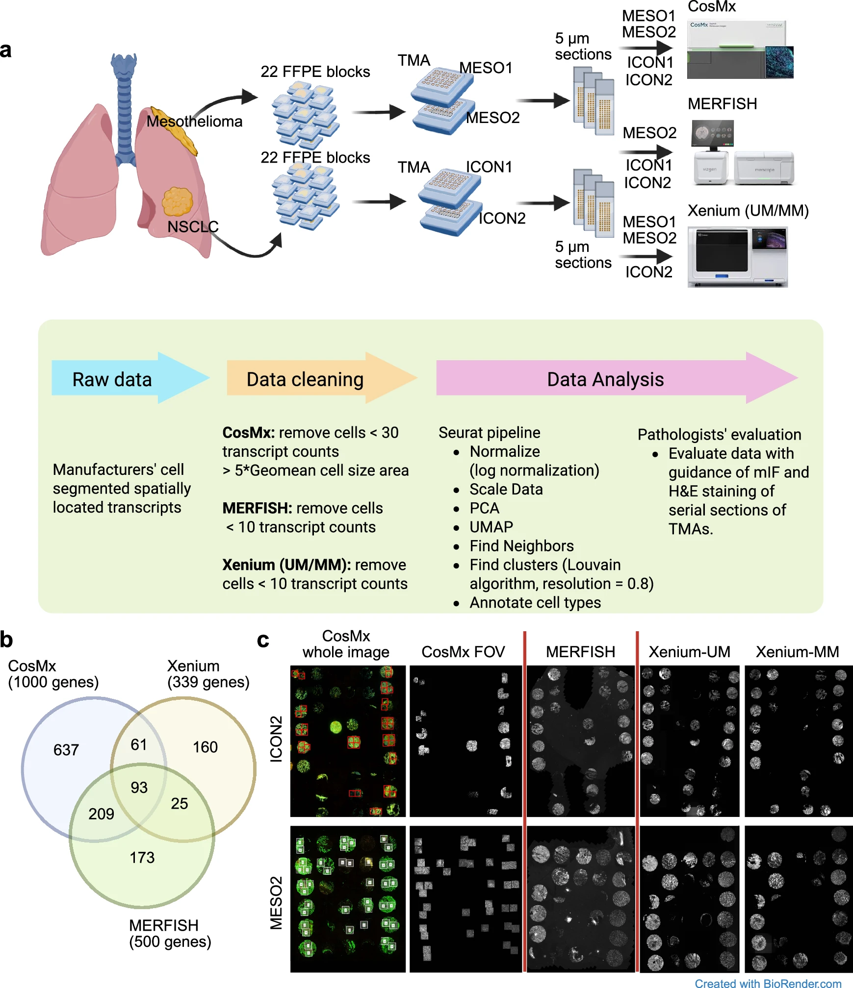
a TMAs containing tissue samples from patients with pleural mesothelioma (MESO1, MESO2; n = 22) or non-small cell lung cancer (NSCLC; ICON1, ICON2; n = 22). Tumor samples were sectioned at a thickness of 5 μm and submitted to CosMx, MERFISH, Xenium-UM, and Xenium-MM assays based on tissue availability. Data analyses were performed using Seurat pipeline and evaluation of pathologists with guidance of mIF and H&E staining. b Venn diagram displays shared genes between panels of the ST platforms. c DAPI staining images of ICON2 and MESO2 TMAs and CosMx whole images of the ICON2 and MESO2 TMAs with staining with morphology markers before Field of View (FOV) selection. Red/white squares display selected FOVs of CosMx. The CosMx FOV, MERFISH, Xenium-UM, and Xenium-MM images display nuclear staining of selected FOV regions and whole tissue cores. Only TMAs with data available from all ST platforms were selected for comparisons
To ensure fair comparisons, the researchers evaluated each platform alongside bulk RNA sequencing, multiplex immunofluorescence, GeoMx spatial profiling, and traditional tissue staining methods. They analyzed automatic and manual cell segmentation and phenotyping to determine how accurately each technology identified different cell types and molecular features within the tumors.
Their results highlighted that each spatial transcriptomics platform has unique strengths and that factors such as probe design can greatly affect data quality. The team also proposed workflows to improve the accuracy and reliability of spatial analyses, offering guidance to researchers choosing the best tool for their cancer studies.
By comparing these technologies head-to-head, the researchers aim to make spatial transcriptomics more robust, standardized, and informative for the growing field of tumor biology.
Ozirmak Lermi N, Molina Ayala M, Hernandez S, Lu W, Khan K, Serrano A, Lubo I, Hamana L, Tomczak K, Barnes S, Dou J, Liang Q, Raso MG, Tang X, Jiang M, Sanchez-Espiridion B, Weissferdt A, Heymach J, Zhang J, Sepesi B, Cascone T, Tsao A, Altan M, Mehran R, Gibbons D, Wistuba I, Haymaker C, Chen K, Solis Soto LM. (2025) Comparison of imaging based single-cell resolution spatial transcriptomics profiling platforms using formalin-fixed paraffin-embedded tumor samples. Nature Communications 16(1):8499. [article]
Related Posts




A research team led by researchers at The University of Texas MD Anderson Cancer Center has taken a closer look at some of the most advanced tools used in spatial transcriptomics, a field that allows scientists to visualize where genes are active within tissues. Understanding how genes are expressed in specific parts of a tumor can reveal critical insights into how cancer develops and interacts with its surrounding microenvironment.
In their work, the team compared three popular imaging-based platforms, CosMx, MERFISH, and Xenium, using lung adenocarcinoma and pleural mesothelioma samples. These technologies can analyze tissues at single-cell resolution, but until now, their strengths and weaknesses had not been systematically compared using the same samples and experimental conditions.
Experimental design, panel comparison, and nuclear staining with the ST platforms
a TMAs containing tissue samples from patients with pleural mesothelioma (MESO1, MESO2; n = 22) or non-small cell lung cancer (NSCLC; ICON1, ICON2; n = 22). Tumor samples were sectioned at a thickness of 5 μm and submitted to CosMx, MERFISH, Xenium-UM, and Xenium-MM assays based on tissue availability. Data analyses were performed using Seurat pipeline and evaluation of pathologists with guidance of mIF and H&E staining. b Venn diagram displays shared genes between panels of the ST platforms. c DAPI staining images of ICON2 and MESO2 TMAs and CosMx whole images of the ICON2 and MESO2 TMAs with staining with morphology markers before Field of View (FOV) selection. Red/white squares display selected FOVs of CosMx. The CosMx FOV, MERFISH, Xenium-UM, and Xenium-MM images display nuclear staining of selected FOV regions and whole tissue cores. Only TMAs with data available from all ST platforms were selected for comparisons
To ensure fair comparisons, the researchers evaluated each platform alongside bulk RNA sequencing, multiplex immunofluorescence, GeoMx spatial profiling, and traditional tissue staining methods. They analyzed automatic and manual cell segmentation and phenotyping to determine how accurately each technology identified different cell types and molecular features within the tumors.
Their results highlighted that each spatial transcriptomics platform has unique strengths and that factors such as probe design can greatly affect data quality. The team also proposed workflows to improve the accuracy and reliability of spatial analyses, offering guidance to researchers choosing the best tool for their cancer studies.
By comparing these technologies head-to-head, the researchers aim to make spatial transcriptomics more robust, standardized, and informative for the growing field of tumor biology.
Ozirmak Lermi N, Molina Ayala M, Hernandez S, Lu W, Khan K, Serrano A, Lubo I, Hamana L, Tomczak K, Barnes S, Dou J, Liang Q, Raso MG, Tang X, Jiang M, Sanchez-Espiridion B, Weissferdt A, Heymach J, Zhang J, Sepesi B, Cascone T, Tsao A, Altan M, Mehran R, Gibbons D, Wistuba I, Haymaker C, Chen K, Solis Soto LM. (2025) Comparison of imaging based single-cell resolution spatial transcriptomics profiling platforms using formalin-fixed paraffin-embedded tumor samples. Nature Communications 16(1):8499. [article]
Related Posts
Recent Posts
 Novel biosensor uses DNA ‘barcodes’ to uncover disease signals in cells
 GP-DHT uses single-cell RNA sequencing to map gene regulatory networks
 Boehringer taps Envisagenics’ AI to mine RNA splicing for cancer targets
 CellART extracts single-cell information from high-resolution spatial transcriptomics
 Cell2space restores spatial context to single-cell RNA sequencing data
 PacBio and TychoBio Collaborate to Train AI Models for Rare Disease
RNA-Seq Products & Services



Stay Connected