

Researchers at Stanford University, have introduced a powerful new computational framework called CytoTRACE 2 that improves our ability to predict how individual cells develop and specialize. This innovation represents a major advance in single-cell biology and data-driven research.
Understanding how cells transition from one type to another, such as how stem cells mature into specialized tissues, has long been a key goal in developmental biology and regenerative medicine. While single-cell RNA sequencing has made it possible to analyze gene expression in thousands of cells at once, interpreting that data to determine each cell’s developmental potential, or ability to become other types of cells, remains a complex challenge.
CytoTRACE 2 uses deep learning, a type of artificial intelligence, to identify molecular features that define a cell’s potential. By analyzing large datasets from multiple tissues and sequencing platforms, the algorithm outperformed earlier models in reconstructing how cells progress through developmental hierarchies.
CytoTRACE 2 architecture and benchmarking metrics
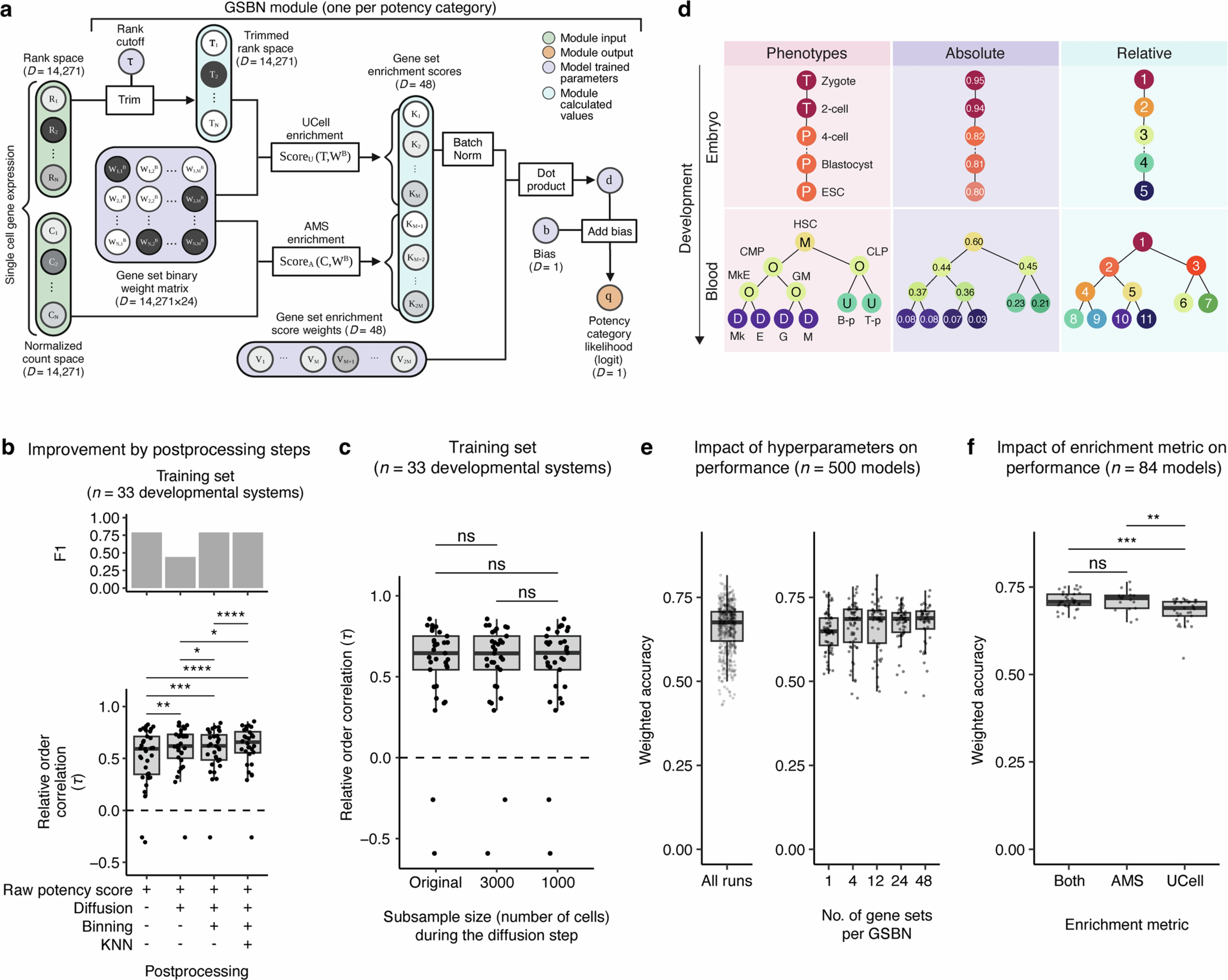
a, Schematic overview of the CytoTRACE 2 core model, focusing on architectural details and key operations between the input layer and output layer for a single gene set binary network (GSBN) module. b, Serial impact of three postprocessing procedures (“Postprocessing” in Methods) on the accuracy of predicting (i) relative developmental orderings (n = 33 systems; bottom) and (ii) potency classes (F1 score, mean-aggregated across six broad potency categories; top) on training datasets (Methods). All results were obtained via leave-one-out cross-validation (LOOCV). c, Box plot showing the performance of CytoTRACE 2 for the prediction of relative orderings after subsampling cells for the Markov diffusion step of the postprocessing procedure (Methods). All training datasets were analyzed using LOOCV d, Illustration of the difference between absolute and relative developmental potential using two hypothetical scRNA-seq datasets, one spanning totipotent through pluripotent cells (‘Embryo’) and the other encompassing multipotent through differentiated cells (‘Blood’). Hypothetical prediction scores are shown for absolute and relative orderings, with the latter reset for each dataset (for example, as for CytoTRACE 1, RNA velocity, Monocle, and CellRank). ESC, embryonic stem cell; HSC, hematopoietic stem cell; CMP, common myeloid progenitor; CLP, common lymphoid progenitor; MkE, megakaryocyte-erythroid progenitor; GM, granulocyte progenitor; B-p, B cell progenitor; T-p, T cell progenitor; Mk, megakaryocyte; E, erythrocyte; G, granulocyte; M, monocyte. e–f, Impact of hyperparameter values on training set performance, showing weighted accuracy for single-cell potency classification using nested LOOCV). e, Left: Each point (n = 500) denotes the results from one iteration of the hyperparameter sweep. Right: Same as the left but showing weighted accuracy plotted by the number of gene sets per GSBN module. f, Weighted accuracy of models trained using distinct gene set enrichment procedures (AMS and/or UCell) after selecting robust hyperparameter values as described in Methods. Statistical significance was determined using a two-sided paired Wilcoxon test in panels b and c and a two-sided unpaired Wilcoxon test in panel f.
The new model provides a clearer, more interpretable view of how cells lose potency as they differentiate, allowing scientists to map developmental landscapes with greater precision. This could accelerate discoveries in areas like stem cell research, cancer biology, and tissue regeneration, where understanding cell fate decisions is essential.
As single-cell RNA sequencing continues to grow in scope and resolution, tools like CytoTRACE 2 will be vital for transforming raw data into meaningful biological insight, helping researchers better understand how cells organize, evolve, and respond to disease.
Availability – R and Python packages for running CytoTRACE 2 with the pre-trained model are freely available for non-profit academic use at https://github.com/digitalcytometry/cytotrace2
Kang M, Gulati GS, Brown EL, Qi Z, Avagyan S, Almagro Armenteros JJ, Gleyzer R, Zhang W, Steen CB, D’Silva JP, Schwab J, Clarke MF, Chaudhuri AA, Newman AM. (2025) Improved reconstruction of single-cell developmental potential with CytoTRACE 2. Nature Methods. [article]
Related Posts


Researchers at Stanford University, have introduced a powerful new computational framework called CytoTRACE 2 that improves our ability to predict how individual cells develop and specialize. This innovation represents a major advance in single-cell biology and data-driven research.
Understanding how cells transition from one type to another, such as how stem cells mature into specialized tissues, has long been a key goal in developmental biology and regenerative medicine. While single-cell RNA sequencing has made it possible to analyze gene expression in thousands of cells at once, interpreting that data to determine each cell’s developmental potential, or ability to become other types of cells, remains a complex challenge.
CytoTRACE 2 uses deep learning, a type of artificial intelligence, to identify molecular features that define a cell’s potential. By analyzing large datasets from multiple tissues and sequencing platforms, the algorithm outperformed earlier models in reconstructing how cells progress through developmental hierarchies.
CytoTRACE 2 architecture and benchmarking metrics
a, Schematic overview of the CytoTRACE 2 core model, focusing on architectural details and key operations between the input layer and output layer for a single gene set binary network (GSBN) module. b, Serial impact of three postprocessing procedures (“Postprocessing” in Methods) on the accuracy of predicting (i) relative developmental orderings (n = 33 systems; bottom) and (ii) potency classes (F1 score, mean-aggregated across six broad potency categories; top) on training datasets (Methods). All results were obtained via leave-one-out cross-validation (LOOCV). c, Box plot showing the performance of CytoTRACE 2 for the prediction of relative orderings after subsampling cells for the Markov diffusion step of the postprocessing procedure (Methods). All training datasets were analyzed using LOOCV d, Illustration of the difference between absolute and relative developmental potential using two hypothetical scRNA-seq datasets, one spanning totipotent through pluripotent cells (‘Embryo’) and the other encompassing multipotent through differentiated cells (‘Blood’). Hypothetical prediction scores are shown for absolute and relative orderings, with the latter reset for each dataset (for example, as for CytoTRACE 1, RNA velocity, Monocle, and CellRank). ESC, embryonic stem cell; HSC, hematopoietic stem cell; CMP, common myeloid progenitor; CLP, common lymphoid progenitor; MkE, megakaryocyte-erythroid progenitor; GM, granulocyte progenitor; B-p, B cell progenitor; T-p, T cell progenitor; Mk, megakaryocyte; E, erythrocyte; G, granulocyte; M, monocyte. e–f, Impact of hyperparameter values on training set performance, showing weighted accuracy for single-cell potency classification using nested LOOCV). e, Left: Each point (n = 500) denotes the results from one iteration of the hyperparameter sweep. Right: Same as the left but showing weighted accuracy plotted by the number of gene sets per GSBN module. f, Weighted accuracy of models trained using distinct gene set enrichment procedures (AMS and/or UCell) after selecting robust hyperparameter values as described in Methods. Statistical significance was determined using a two-sided paired Wilcoxon test in panels b and c and a two-sided unpaired Wilcoxon test in panel f.
The new model provides a clearer, more interpretable view of how cells lose potency as they differentiate, allowing scientists to map developmental landscapes with greater precision. This could accelerate discoveries in areas like stem cell research, cancer biology, and tissue regeneration, where understanding cell fate decisions is essential.
As single-cell RNA sequencing continues to grow in scope and resolution, tools like CytoTRACE 2 will be vital for transforming raw data into meaningful biological insight, helping researchers better understand how cells organize, evolve, and respond to disease.
Availability – R and Python packages for running CytoTRACE 2 with the pre-trained model are freely available for non-profit academic use at https://github.com/digitalcytometry/cytotrace2
Kang M, Gulati GS, Brown EL, Qi Z, Avagyan S, Almagro Armenteros JJ, Gleyzer R, Zhang W, Steen CB, D’Silva JP, Schwab J, Clarke MF, Chaudhuri AA, Newman AM. (2025) Improved reconstruction of single-cell developmental potential with CytoTRACE 2. Nature Methods. [article]
Related Posts
Recent Posts
 RNAmel simplifies RNA sequencing data analysis with a reproducible, integrated platform
 Heart failure reshapes bone marrow to create inflammation-related immune memory
 Overlooked DNA regions surrounding ‘microexons’ hold missing pieces of autism’s genetic puzzle
 RiboBA improves detection of hidden protein-coding regions using ribosome profiling
 An anti-senescence gene may fight spinal disc deterioration
 Bulk RNA-seq Analysis: A Complete Beginner’s Guide
RNA-Seq Products & Services


Stay Connected