

Researchers at Yale University have developed a new approach called DBiTplus, short for Deterministic Barcoding in Tissue sequencing plus, that overcomes this limitation. DBiTplus allows scientists to measure RNA and proteins from the same tissue section, creating a detailed spatial map of both the transcriptome and proteome.
The method works by applying a grid of molecular barcodes directly onto intact tissue. These barcodes label RNA molecules in defined spatial locations, enabling RNA sequencing data to be traced back to their exact positions in the tissue. After RNA capture, the tissue remains intact, allowing the same section to be stained and imaged for dozens of proteins using multiplexed imaging techniques. This means gene expression and protein patterns can be directly compared cell by cell.
DBiTplus workflow and overview of bioinformatic workflow for integrative multimodal data analysis
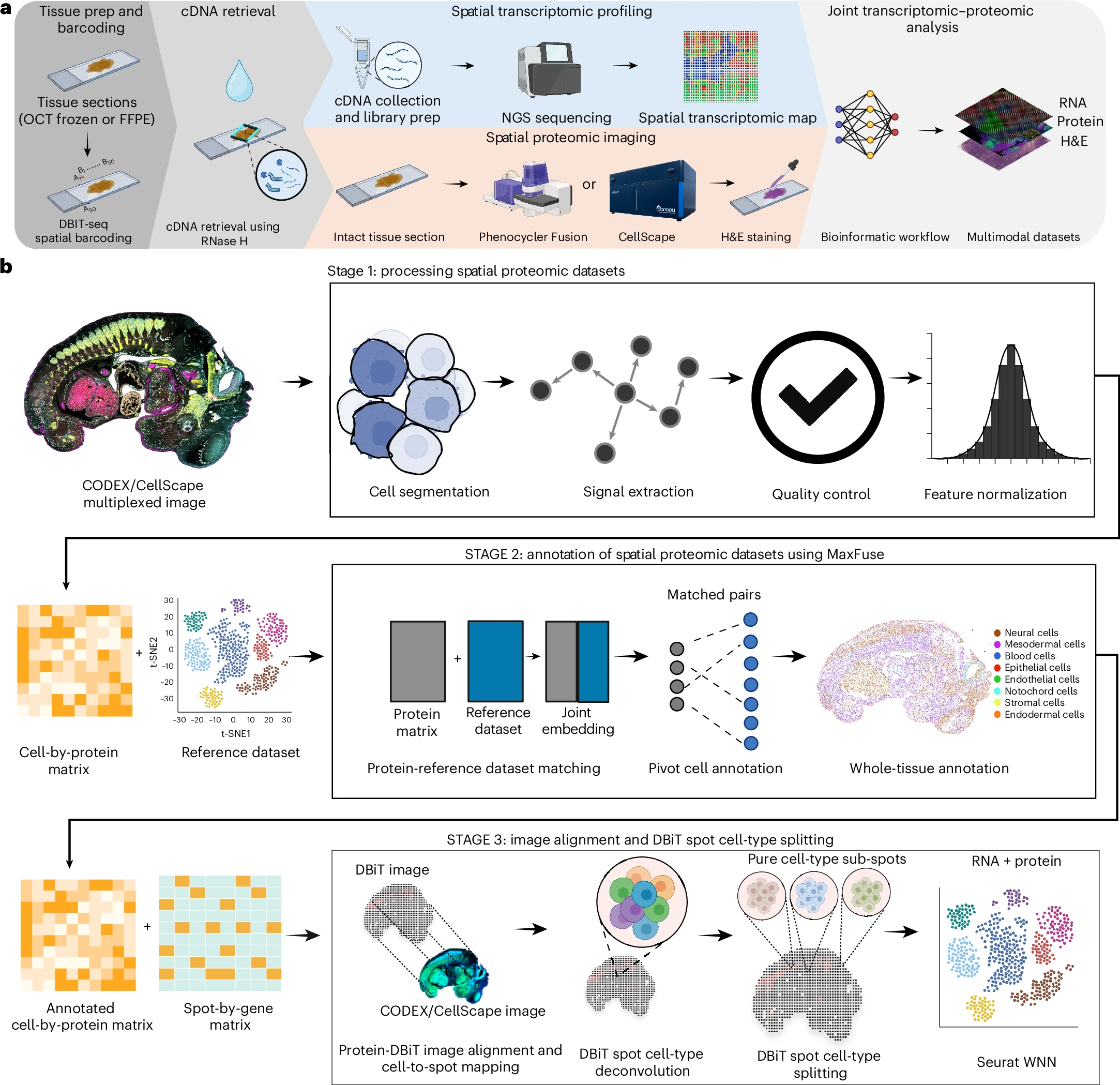
a, Workflow of DBiTplus technology. b, The three steps of the integrative analysis. DBIT-seq, Deterministic Barcoding in Tissue sequencing; NGS, next-generation sequencing.
To make sense of these rich datasets, the team developed computational pipelines that integrate imaging and sequencing data. Protein imaging guides the identification of individual cells, while RNA sequencing provides transcriptome-wide information about gene activity within each cell. Together, this creates single-cell resolved spatial atlases that reveal how cells are organized and how biological pathways operate within real tissue environments.
The researchers demonstrated DBiTplus in a range of challenging samples, including mouse embryos and formalin-fixed paraffin-embedded human lymph nodes and lymphoma tissues. Importantly, the approach uncovered spatial gene expression patterns linked to lymphoma development and progression, showing how cancer cells and their surrounding microenvironment interact at a molecular level.
By combining spatial RNA sequencing and protein imaging in one unified workflow, DBiTplus provides a powerful way to study tissue biology, disease mechanisms, and clinical samples that were previously difficult to analyze in depth.
Availability – The code for the analyzing the processed mxIF imaging datasets and the sequencing datasets are available on Zenodo (https://doi.org/10.5281/zenodo.16827046) and GitHub (https://github.com/Janezjz/DBiT-plus/).
Enninful A, Zhang Z, Klymyshyn D, Ingalls M, Yang M, Zong H, Bai Z, Farzad N, Su G, Baysoy A, Nam J, Lu Y, Bao S, Deng S, Zhang NR, Braubach O, Xu ML, Ma Z, Fan R. (2026) Integration of imaging-based and sequencing-based spatial omics mapping on the same tissue section via DBiTplus. Nature Methods [Epub ahead of print]. [article]
Related Posts






Researchers at Yale University have developed a new approach called DBiTplus, short for Deterministic Barcoding in Tissue sequencing plus, that overcomes this limitation. DBiTplus allows scientists to measure RNA and proteins from the same tissue section, creating a detailed spatial map of both the transcriptome and proteome.
The method works by applying a grid of molecular barcodes directly onto intact tissue. These barcodes label RNA molecules in defined spatial locations, enabling RNA sequencing data to be traced back to their exact positions in the tissue. After RNA capture, the tissue remains intact, allowing the same section to be stained and imaged for dozens of proteins using multiplexed imaging techniques. This means gene expression and protein patterns can be directly compared cell by cell.
DBiTplus workflow and overview of bioinformatic workflow for integrative multimodal data analysis
a, Workflow of DBiTplus technology. b, The three steps of the integrative analysis. DBIT-seq, Deterministic Barcoding in Tissue sequencing; NGS, next-generation sequencing.
To make sense of these rich datasets, the team developed computational pipelines that integrate imaging and sequencing data. Protein imaging guides the identification of individual cells, while RNA sequencing provides transcriptome-wide information about gene activity within each cell. Together, this creates single-cell resolved spatial atlases that reveal how cells are organized and how biological pathways operate within real tissue environments.
The researchers demonstrated DBiTplus in a range of challenging samples, including mouse embryos and formalin-fixed paraffin-embedded human lymph nodes and lymphoma tissues. Importantly, the approach uncovered spatial gene expression patterns linked to lymphoma development and progression, showing how cancer cells and their surrounding microenvironment interact at a molecular level.
By combining spatial RNA sequencing and protein imaging in one unified workflow, DBiTplus provides a powerful way to study tissue biology, disease mechanisms, and clinical samples that were previously difficult to analyze in depth.
Availability – The code for the analyzing the processed mxIF imaging datasets and the sequencing datasets are available on Zenodo (https://doi.org/10.5281/zenodo.16827046) and GitHub (https://github.com/Janezjz/DBiT-plus/).
Enninful A, Zhang Z, Klymyshyn D, Ingalls M, Yang M, Zong H, Bai Z, Farzad N, Su G, Baysoy A, Nam J, Lu Y, Bao S, Deng S, Zhang NR, Braubach O, Xu ML, Ma Z, Fan R. (2026) Integration of imaging-based and sequencing-based spatial omics mapping on the same tissue section via DBiTplus. Nature Methods [Epub ahead of print]. [article]
Related Posts
Recent Posts
 Scientists identify the neuroblastoma cells that signal poor outcomes
 GARAGE improves rare cell detection in single-cell RNA sequencing analysis using AI
 Machine learning reveals how RNA splicing is regulated in individual cell types
 Illumina releases SpliceAI2 to help advance rare disease research
 Postdoctoral Fellow – Long-Read RNA Methods Development for Neurodegeneration
 Novel biosensor uses DNA ‘barcodes’ to uncover disease signals in cells
RNA-Seq Products & Services


Stay Connected