

Three-dimensional (3D) structure of DNA in our cells changes across different tissues. The study of these chromatin structures—how DNA is packaged within the cell—has revealed important insights into how cells function. However, previous methods have had limitations, especially when it comes to studying a large number of cells at once.
Researchers at the Peking University have developed a groundbreaking approach: the high-throughput single-cell Hi-C (dscHi-C) method, combined with its multi-omics version, dscHi-C-multiome. This advanced technique not only looks at the 3D structure of the genome in individual cells but also allows scientists to study gene expression (the transcriptome) in the same cell, providing a much richer understanding of cellular behavior.
Droplet-based single-cell joint Hi-C and gene expression co-assay
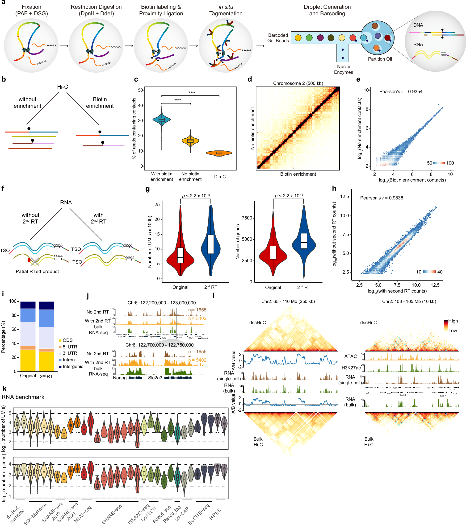
a Schematics of the droplet single-cell simultaneous Hi-C and RNA profiling method. b Schematic showing two Hi-C part processing procedures, with and without biotin enrichment. c Violin plot comparing the percentage of reads containing contacts for with and without biotin enrichment, Dip-C was plotted for comparison. d Contact map of with (bottom right) and without (top left) biotin enrichment of chromosome 2 at 500-kb resolution. e Density plot showing the correlation of raw Hi-C contact counts at 500-kb resolution between with and without biotin enrichment. f Schematic showing two RNA part processing procedures, with and without second RT. g Violin plot comparing the scRNA-seq library size (left) and the number of genes (right) with and without second RT. h Density plot showing the correlation of raw RNA counts for each gene between with and without second RT. i RNA reads the composition of with and without second RT RNA libraries. j Aggregated scRNA-seq profiles along with bulk RNA-seq of mESC at Nanog genomic loci. k Benchmark of scRNA-seq library size (top) and the number of detected genes (bottom) between dscHi-C multiome and other published single-cell multi-omics datasets. l Contact maps of ensemble dscHi-C multiome and bulk Hi-C accompanied with eigenvalue, RNA-seq track, ATAC-seq, and H3K27ac tracks.
In the context of aging, researchers used this dscHi-C-multiome approach to study brain cells in mice across three different ages (3, 12, and 23 months). By analyzing more than 32,000 individual cells, they were able to map out how the 3D genome architecture changes over time and how these changes correlate with gene expression.
One of the standout features of the dscHi-C-multiome technique is its ability to identify specific cell types in the adult mouse brain while simultaneously measuring both the physical structure of the genome and the expression of genes. This “multi-omics” approach is like getting two pieces of the puzzle at once. By combining structural data with gene expression data, scientists can uncover relationships between the organization of the genome and how genes are turned on or off in response to aging.
The results of this study highlighted key changes in specific cell types. For example, in neurons, the changes in genome structure were related to metabolic processes and cell shape changes, while in glial cells, the changes were linked to immune responses. These findings show that 3D genome organization plays a significant role in how the brain ages and how its cells adapt over time.
In short, the dscHi-C-multiome method is a powerful tool that allows scientists to dive deeper into the world of genome architecture and gene expression. It opens the door to large-scale studies that can explore how our cells and tissues change during development, aging, and disease, providing valuable insights for future research in neuroscience and beyond.
Wu H, Wang M, Zheng Y, Xie XS. (2025) Droplet-based high-throughput 3D genome structure mapping of single cells with simultaneous transcriptomics. Cell Discov 11(1):8. [article]
Related Posts






Three-dimensional (3D) structure of DNA in our cells changes across different tissues. The study of these chromatin structures—how DNA is packaged within the cell—has revealed important insights into how cells function. However, previous methods have had limitations, especially when it comes to studying a large number of cells at once.
Researchers at the Peking University have developed a groundbreaking approach: the high-throughput single-cell Hi-C (dscHi-C) method, combined with its multi-omics version, dscHi-C-multiome. This advanced technique not only looks at the 3D structure of the genome in individual cells but also allows scientists to study gene expression (the transcriptome) in the same cell, providing a much richer understanding of cellular behavior.
Droplet-based single-cell joint Hi-C and gene expression co-assay
a Schematics of the droplet single-cell simultaneous Hi-C and RNA profiling method. b Schematic showing two Hi-C part processing procedures, with and without biotin enrichment. c Violin plot comparing the percentage of reads containing contacts for with and without biotin enrichment, Dip-C was plotted for comparison. d Contact map of with (bottom right) and without (top left) biotin enrichment of chromosome 2 at 500-kb resolution. e Density plot showing the correlation of raw Hi-C contact counts at 500-kb resolution between with and without biotin enrichment. f Schematic showing two RNA part processing procedures, with and without second RT. g Violin plot comparing the scRNA-seq library size (left) and the number of genes (right) with and without second RT. h Density plot showing the correlation of raw RNA counts for each gene between with and without second RT. i RNA reads the composition of with and without second RT RNA libraries. j Aggregated scRNA-seq profiles along with bulk RNA-seq of mESC at Nanog genomic loci. k Benchmark of scRNA-seq library size (top) and the number of detected genes (bottom) between dscHi-C multiome and other published single-cell multi-omics datasets. l Contact maps of ensemble dscHi-C multiome and bulk Hi-C accompanied with eigenvalue, RNA-seq track, ATAC-seq, and H3K27ac tracks.
In the context of aging, researchers used this dscHi-C-multiome approach to study brain cells in mice across three different ages (3, 12, and 23 months). By analyzing more than 32,000 individual cells, they were able to map out how the 3D genome architecture changes over time and how these changes correlate with gene expression.
One of the standout features of the dscHi-C-multiome technique is its ability to identify specific cell types in the adult mouse brain while simultaneously measuring both the physical structure of the genome and the expression of genes. This “multi-omics” approach is like getting two pieces of the puzzle at once. By combining structural data with gene expression data, scientists can uncover relationships between the organization of the genome and how genes are turned on or off in response to aging.
The results of this study highlighted key changes in specific cell types. For example, in neurons, the changes in genome structure were related to metabolic processes and cell shape changes, while in glial cells, the changes were linked to immune responses. These findings show that 3D genome organization plays a significant role in how the brain ages and how its cells adapt over time.
In short, the dscHi-C-multiome method is a powerful tool that allows scientists to dive deeper into the world of genome architecture and gene expression. It opens the door to large-scale studies that can explore how our cells and tissues change during development, aging, and disease, providing valuable insights for future research in neuroscience and beyond.
Wu H, Wang M, Zheng Y, Xie XS. (2025) Droplet-based high-throughput 3D genome structure mapping of single cells with simultaneous transcriptomics. Cell Discov 11(1):8. [article]
Related Posts
Recent Posts
 Machine learning reveals how RNA splicing is regulated in individual cell types
 Illumina releases SpliceAI2 to help advance rare disease research
 Postdoctoral Fellow – Long-Read RNA Methods Development for Neurodegeneration
 Novel biosensor uses DNA ‘barcodes’ to uncover disease signals in cells
 GP-DHT uses single-cell RNA sequencing to map gene regulatory networks
 Boehringer taps Envisagenics’ AI to mine RNA splicing for cancer targets
RNA-Seq Products & Services


Stay Connected