

Researchers from Baylor College of Medicine have uncovered a previously unknown way that DNA damage occurs during transcription—when genes are being read to produce RNA. This discovery adds a fourth mechanism to the known ways that transcription can harm DNA, shedding light on potential contributors to cancer, aging, and neurodegenerative diseases.
Understanding DNA Damage During Transcription
Cells rely on DNA as their instruction manual, but over time, damage to this genetic code can lead to mutations. Some of these mutations may cause cells to grow uncontrollably (as in cancer) or contribute to age-related diseases. Scientists have long known that DNA damage can occur when the molecular machinery that reads genes (RNA polymerase) collides with the machinery that copies DNA (the replication complex). Another known cause is R-loops, structures formed when RNA gets tangled with DNA, leading to breaks.
Now, these researchers have found a fourth source of damage: when transcription ends, single-stranded DNA (ssDNA) is left vulnerable. Their study reveals that these single-stranded DNA ends, particularly at specific termination sequences, can become hotspots for DNA damage and genomic instability.
How Did the Researchers Find This?
To track where DNA damage occurs, the team engineered special proteins that can detect and capture exposed single-stranded DNA ends. They tested this system in both bacteria (E. coli) and human cells, and to their surprise, they found that DNA damage at transcription termination sites was much more common than expected—at least once per cell division in bacteria.
The researchers also developed a technique called ThreeSSeq, which allowed them to map these DNA damage hotspots across the genome. They found that DNA damage occurred frequently near sequences resembling a transcription stop signal (5’-CCTTTTTT-3’). These hotspots coincided with RNA 3’-end sites identified by RNA sequencing methods, such as DirectRNA sequencing and SEnd-seq. Importantly, when the team modified RNA polymerase to ignore transcription stop signals, the damage was reduced, suggesting that the transcription termination process itself contributes to the instability.
ThreeSSeq 3’-ssDNA-end genomic profiling with DExI detects dispersed, reproducible, hotspots
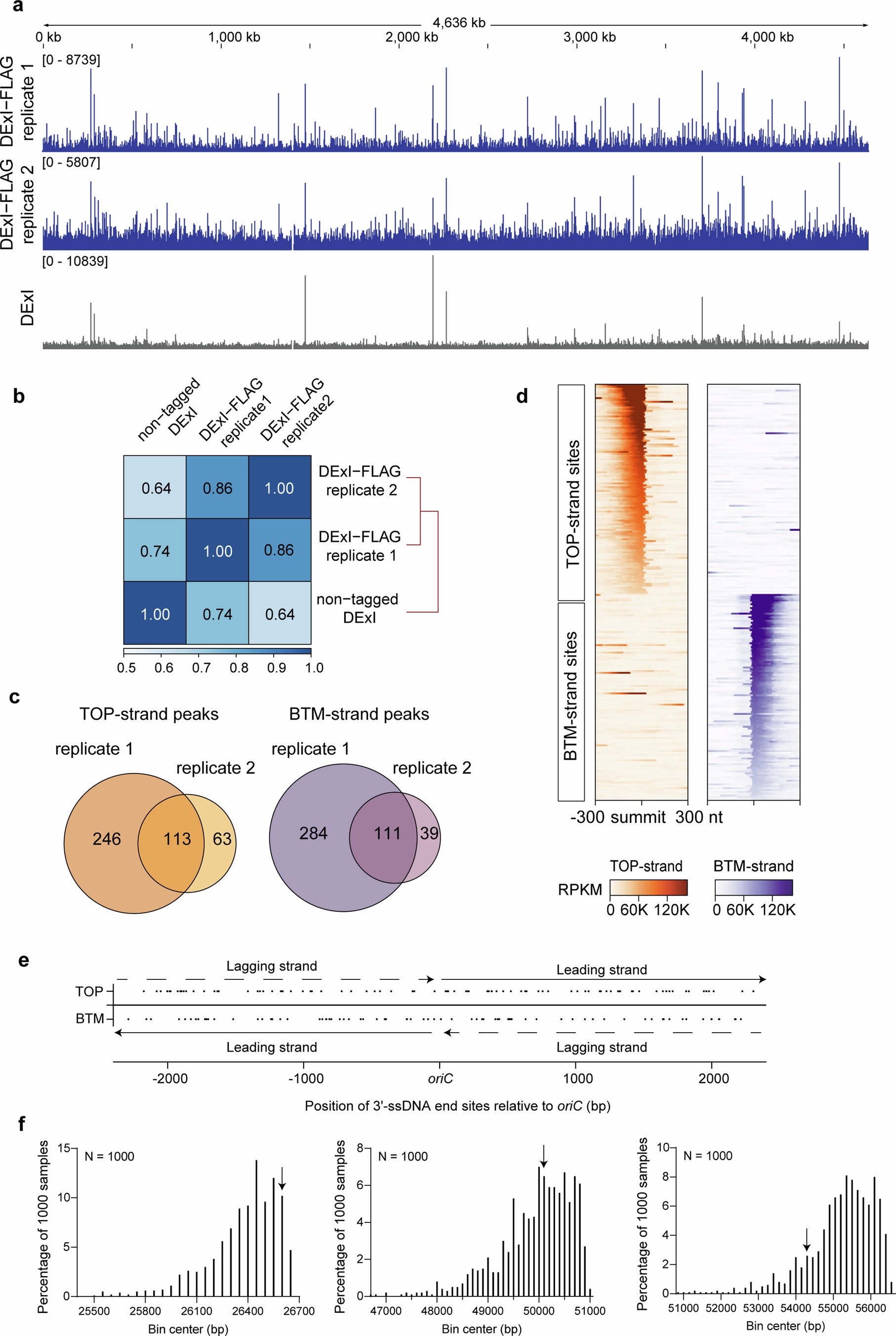
a, Integrated genome viewer (IGV) views of signals from ThreeSSeq with DExI in the indicated strains reveal hotspots throughout the genome. Signals were normalized by Reads Per Kilobase per Million mapped reads (RPKM). For calling of peaks and all quantification, the DExI signal (negative control, background) is subtracted from the signal with DExI-FLAG to obtain the DExI-FLAG-specific reads. b, Two DExI-FLAG experiments are more correlated with each other than with the DExI control. Heatmap of Pearson correlations between the experiments. c, Venn diagrams of peak overlap between two replicates in DExI-FLAG-producing, otherwise wild-type cells. d, Heatmaps of ThreeSSeq signals within ± 300 nt regions around peak summits of 113 TOP-strand and 111 BTM-strand 3’-ssDNA-end hotspot sites. Sites were centered along peak summits and sorted by signal intensity. Signals were normalized by RPKM. e, The distribution of 175 sequence-specific 3’-ssDNA-end sites (shown as black dots) throughout the genome. The leading- and lagging-strand in each replichore are indicated. f, Sequence-specific 3’-ssDNA-end hotspots are distributed randomly throughout the genome. To test the randomness of the 3’-ssDNA-end-hotspot distribution, we sampled 175, 92 and 83 genomic sites, corresponding to the number of all DExI-FLAG-immunoprecipitated sites throughout the genome, the number of TOP-strand sites and the number of BTM-strand sites, randomly 1000 times, and calculated the average distance of neighboring sites. The barplots display the distribution of the sample means, and the arrows indicate the average distances of tested data, including all 175 sites (left, p = 0.10), 92 TOP-strand sites (center, p = 0.48), and 83 BTM-strand sites (right, p = 0.16). The distribution of actual 3’-ssDNA-end hotspots was not different from the 1000 random samples. Strains used: a: SMR24776, SMR24777; b: SMR24776, SMR24777; d: SMR24776.
These findings provide a new perspective on how cells accumulate DNA damage over time, which could have significant implications for cancer research and age-related diseases. By understanding how transcription termination leaves DNA vulnerable, scientists may be able to develop strategies to prevent or repair this type of damage, ultimately reducing the risk of mutations that drive disease.
Availability – The main scripts used for the analysis are available from GitHub (https://github.com/JLiu0504/ThreeSSeq-processing).
Liu J, Perren JO, Rogers CM, Nimer S, Wen AX, Halliday JA, Fitzgerald DM, Mei Q, Nehring RB, Crum M, Kozmin SG, Xia J, Cooke MB, Zhai Y, Bates D, Li L, Hastings PJ, Artsimovitch I, Herman C, Sung PM, Miller KM, Rosenberg SM. (2025) Endogenous DNA damage at sites of terminated transcripts. Nature. [Epub ahead of print]. [article]
Related Posts




Researchers from Baylor College of Medicine have uncovered a previously unknown way that DNA damage occurs during transcription—when genes are being read to produce RNA. This discovery adds a fourth mechanism to the known ways that transcription can harm DNA, shedding light on potential contributors to cancer, aging, and neurodegenerative diseases.
Understanding DNA Damage During Transcription
Cells rely on DNA as their instruction manual, but over time, damage to this genetic code can lead to mutations. Some of these mutations may cause cells to grow uncontrollably (as in cancer) or contribute to age-related diseases. Scientists have long known that DNA damage can occur when the molecular machinery that reads genes (RNA polymerase) collides with the machinery that copies DNA (the replication complex). Another known cause is R-loops, structures formed when RNA gets tangled with DNA, leading to breaks.
Now, these researchers have found a fourth source of damage: when transcription ends, single-stranded DNA (ssDNA) is left vulnerable. Their study reveals that these single-stranded DNA ends, particularly at specific termination sequences, can become hotspots for DNA damage and genomic instability.
How Did the Researchers Find This?
To track where DNA damage occurs, the team engineered special proteins that can detect and capture exposed single-stranded DNA ends. They tested this system in both bacteria (E. coli) and human cells, and to their surprise, they found that DNA damage at transcription termination sites was much more common than expected—at least once per cell division in bacteria.
The researchers also developed a technique called ThreeSSeq, which allowed them to map these DNA damage hotspots across the genome. They found that DNA damage occurred frequently near sequences resembling a transcription stop signal (5’-CCTTTTTT-3’). These hotspots coincided with RNA 3’-end sites identified by RNA sequencing methods, such as DirectRNA sequencing and SEnd-seq. Importantly, when the team modified RNA polymerase to ignore transcription stop signals, the damage was reduced, suggesting that the transcription termination process itself contributes to the instability.
ThreeSSeq 3’-ssDNA-end genomic profiling with DExI detects dispersed, reproducible, hotspots
a, Integrated genome viewer (IGV) views of signals from ThreeSSeq with DExI in the indicated strains reveal hotspots throughout the genome. Signals were normalized by Reads Per Kilobase per Million mapped reads (RPKM). For calling of peaks and all quantification, the DExI signal (negative control, background) is subtracted from the signal with DExI-FLAG to obtain the DExI-FLAG-specific reads. b, Two DExI-FLAG experiments are more correlated with each other than with the DExI control. Heatmap of Pearson correlations between the experiments. c, Venn diagrams of peak overlap between two replicates in DExI-FLAG-producing, otherwise wild-type cells. d, Heatmaps of ThreeSSeq signals within ± 300 nt regions around peak summits of 113 TOP-strand and 111 BTM-strand 3’-ssDNA-end hotspot sites. Sites were centered along peak summits and sorted by signal intensity. Signals were normalized by RPKM. e, The distribution of 175 sequence-specific 3’-ssDNA-end sites (shown as black dots) throughout the genome. The leading- and lagging-strand in each replichore are indicated. f, Sequence-specific 3’-ssDNA-end hotspots are distributed randomly throughout the genome. To test the randomness of the 3’-ssDNA-end-hotspot distribution, we sampled 175, 92 and 83 genomic sites, corresponding to the number of all DExI-FLAG-immunoprecipitated sites throughout the genome, the number of TOP-strand sites and the number of BTM-strand sites, randomly 1000 times, and calculated the average distance of neighboring sites. The barplots display the distribution of the sample means, and the arrows indicate the average distances of tested data, including all 175 sites (left, p = 0.10), 92 TOP-strand sites (center, p = 0.48), and 83 BTM-strand sites (right, p = 0.16). The distribution of actual 3’-ssDNA-end hotspots was not different from the 1000 random samples. Strains used: a: SMR24776, SMR24777; b: SMR24776, SMR24777; d: SMR24776.
These findings provide a new perspective on how cells accumulate DNA damage over time, which could have significant implications for cancer research and age-related diseases. By understanding how transcription termination leaves DNA vulnerable, scientists may be able to develop strategies to prevent or repair this type of damage, ultimately reducing the risk of mutations that drive disease.
Availability – The main scripts used for the analysis are available from GitHub (https://github.com/JLiu0504/ThreeSSeq-processing).
Liu J, Perren JO, Rogers CM, Nimer S, Wen AX, Halliday JA, Fitzgerald DM, Mei Q, Nehring RB, Crum M, Kozmin SG, Xia J, Cooke MB, Zhai Y, Bates D, Li L, Hastings PJ, Artsimovitch I, Herman C, Sung PM, Miller KM, Rosenberg SM. (2025) Endogenous DNA damage at sites of terminated transcripts. Nature. [Epub ahead of print]. [article]
Related Posts
Recent Posts
 Scientists identify the neuroblastoma cells that signal poor outcomes
 GARAGE improves rare cell detection in single-cell RNA sequencing analysis using AI
 Machine learning reveals how RNA splicing is regulated in individual cell types
 Illumina releases SpliceAI2 to help advance rare disease research
 Postdoctoral Fellow – Long-Read RNA Methods Development for Neurodegeneration
 Novel biosensor uses DNA ‘barcodes’ to uncover disease signals in cells
RNA-Seq Products & Services



Stay Connected