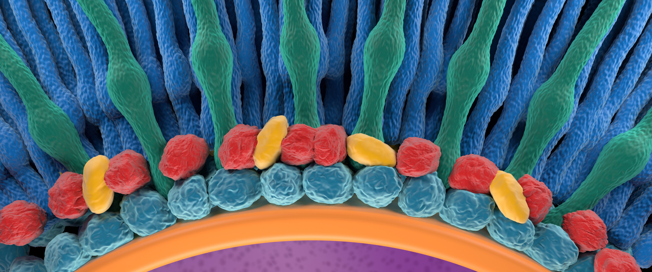
By discovering retinal cell subgroups in mice, researchers hope to pinpoint an optimal cell type for transplantation to restore vision in blinding conditions.
A new understanding of retinal cell development may help pave the way for future retina transplants, which could restore sight to people whose conditions currently have no effective treatments, according to researchers at the Perelman School of Medicine at the University of Pennsylvania. Their findings were published today in Frontiers in Cell and Developmental Biology.
Currently, the transplantation of healthy photoreceptor cells—neurons that detect light in the retina at the back of the eye—is largely ineffective because very few of the donated cells successfully connect to restore vision. However, the treatment remains promising if an ideal cell population that is more likely to form connections could be found.
By identifying three different developmental stages of photoreceptor cells in mice—which the study group believes may have analogous cell populations in humans—the researchers hope to determine which cell population is most capable of forming neuronal connections in the eye. Their findings may help researchers identify which cells are most likely to survive transplantation and integrate into the eye, leading to improved treatment outcomes.
“We plan to isolate and transplant each subgroup individually, in the hopes that transplanting a more pure cell population will improve future cell-based therapies to improve vision in late-stage blinding conditions,” said the study’s principal investigator, Katherine Uyhazi, MD, PhD, an assistant professor of Ophthalmology.
Differences in cells and the possibilities they contain
Retinal diseases are a leading cause of blindness, affecting millions of people worldwide. Many of these conditions are inherited, while others, such as age-related macular degeneration, do not have a known genetic cause. Current treatments are aimed at slowing or stopping vision loss, but cell-based therapies have the potential to reverse vision loss by replacing lost or damaged cells.
“We know that retinal development does not occur uniformly, but in waves,” Uyhazi said. “There is a mix of developmental cell stages present at any given chronological age during retinal development.” Learning more about retinal development presented an opportunity to optimize retinal regeneration in disease.
Which is which
By using single cell RNA sequencing, which can analyze how genes are expressed in individual cells, first author Joseph Yano, a PhD candidate in Uyhazi’s lab, was able to identify three distinct states of developing photoreceptor cells: early, mid, and late.
The team further showed that there may be comparable populations of cells in human retinal organoids, which are three-dimensional lab-grown structures that mimic structures in the human eye.
What’s happening now
Following up on their discoveries, Uyhazi and her team are now working on methods for isolating and transplanting each of these cell states in the retina. Early cells are more similar to stem cells and may better survive transplantation, while late cells are more similar to retinal cells that can respond to light.
Through these further experiments in mice, the team hopes to identify a “goldilocks” stage of cell that is best suited to help repair the retina.
Source – Penn Medicine


By discovering retinal cell subgroups in mice, researchers hope to pinpoint an optimal cell type for transplantation to restore vision in blinding conditions.
A new understanding of retinal cell development may help pave the way for future retina transplants, which could restore sight to people whose conditions currently have no effective treatments, according to researchers at the Perelman School of Medicine at the University of Pennsylvania. Their findings were published today in Frontiers in Cell and Developmental Biology.
Currently, the transplantation of healthy photoreceptor cells—neurons that detect light in the retina at the back of the eye—is largely ineffective because very few of the donated cells successfully connect to restore vision. However, the treatment remains promising if an ideal cell population that is more likely to form connections could be found.
By identifying three different developmental stages of photoreceptor cells in mice—which the study group believes may have analogous cell populations in humans—the researchers hope to determine which cell population is most capable of forming neuronal connections in the eye. Their findings may help researchers identify which cells are most likely to survive transplantation and integrate into the eye, leading to improved treatment outcomes.
Differences in cells and the possibilities they contain
Retinal diseases are a leading cause of blindness, affecting millions of people worldwide. Many of these conditions are inherited, while others, such as age-related macular degeneration, do not have a known genetic cause. Current treatments are aimed at slowing or stopping vision loss, but cell-based therapies have the potential to reverse vision loss by replacing lost or damaged cells.
Which is which
By using single cell RNA sequencing, which can analyze how genes are expressed in individual cells, first author Joseph Yano, a PhD candidate in Uyhazi’s lab, was able to identify three distinct states of developing photoreceptor cells: early, mid, and late.
The team further showed that there may be comparable populations of cells in human retinal organoids, which are three-dimensional lab-grown structures that mimic structures in the human eye.
What’s happening now
Following up on their discoveries, Uyhazi and her team are now working on methods for isolating and transplanting each of these cell states in the retina. Early cells are more similar to stem cells and may better survive transplantation, while late cells are more similar to retinal cells that can respond to light.
Through these further experiments in mice, the team hopes to identify a “goldilocks” stage of cell that is best suited to help repair the retina.
Source – Penn Medicine
Yano JJ, Wei Z, Gajjar KJ, Barati-Stec KT, Yang E, Uyhazi KE. (2026) Lineage tracing reveals photoreceptor precursor cell subpopulations that contribute to murine retinogenesis. Frontiers in Cell and Developmental Biology 14:1814134. [article]
Related Posts
Novel biosensor uses DNA ‘barcodes’ to uncover disease signals in cells
CellART extracts single-cell information from high-resolution spatial transcriptomics
Cell2space restores spatial context to single-cell RNA sequencing data
Urine test may improve bladder cancer diagnosis and guide treatment decisions
DeCoRE – a computational method to resolve RNA structural heterogeneity from RNA structure probing data by direct RNA sequencing
Tracing 700 million years of cell evolution
How should biological replicates be handled in long-read RNA sequencing?
RNA-Seq reveals a new way the developing heart controls its growth
The Human RNome Project Consortium seeks to publish first-ever complete sequence of the body’s RNA
scFlowReport brings multiple single-cell RNA sequencing analyses into one workflow
Long-read RNA sequencing reveals how neurons choose alternative RNA endings
DeOPUS improves cell-type estimates from bulk RNA sequencing
ResolVI improves accuracy in spatial transcriptomics analysis
RNA sequencing reveals transcriptional allele differences in prostate cancer stem cells
Unmasking “zombie cells” in aging tissue with an AI-powered barcode
RNA variant imaging for cancer diagnosis
Autism-risk mutations reveal two opposing patterns of brain gene activity
A real-data approach to evaluating RNA transcript analysis methods
Detection and sequencing of Ap2N-capped RNAs in human cells
Single-cell RNA sequencing provides a closer look at the Aedes aegypti midgut
Stay Connected
Submit a Post to the Blog
Recent Posts
Subscribe to the RNA-Seq Blog
RNA-Seq Products & Services