The maternal microbiome and immune system have both independent and synergistic effects on fetal brain health, changes in the mother’s immune system have been linked to an increased risk of neurodevelopmental disorders in children. A new study, published today in Nature Neuroscience, expands our understanding of this “gut-immune axis” by mapping the impact of stressors during pregnancy, namely changes in the microbiome and activation of the immune system, on the neuroimmune landscape of the developing fetal brain.
The research team, from the Division of Newborn Medicine at Boston Children’s Hospital and the SickKids Research Institute, have discovered notable sex-specific responses, including vulnerability for a specific immune pathway in the male brain that could be a potential target for early intervention.
MERFISH of the embryonic mouse brain at E14 and E18
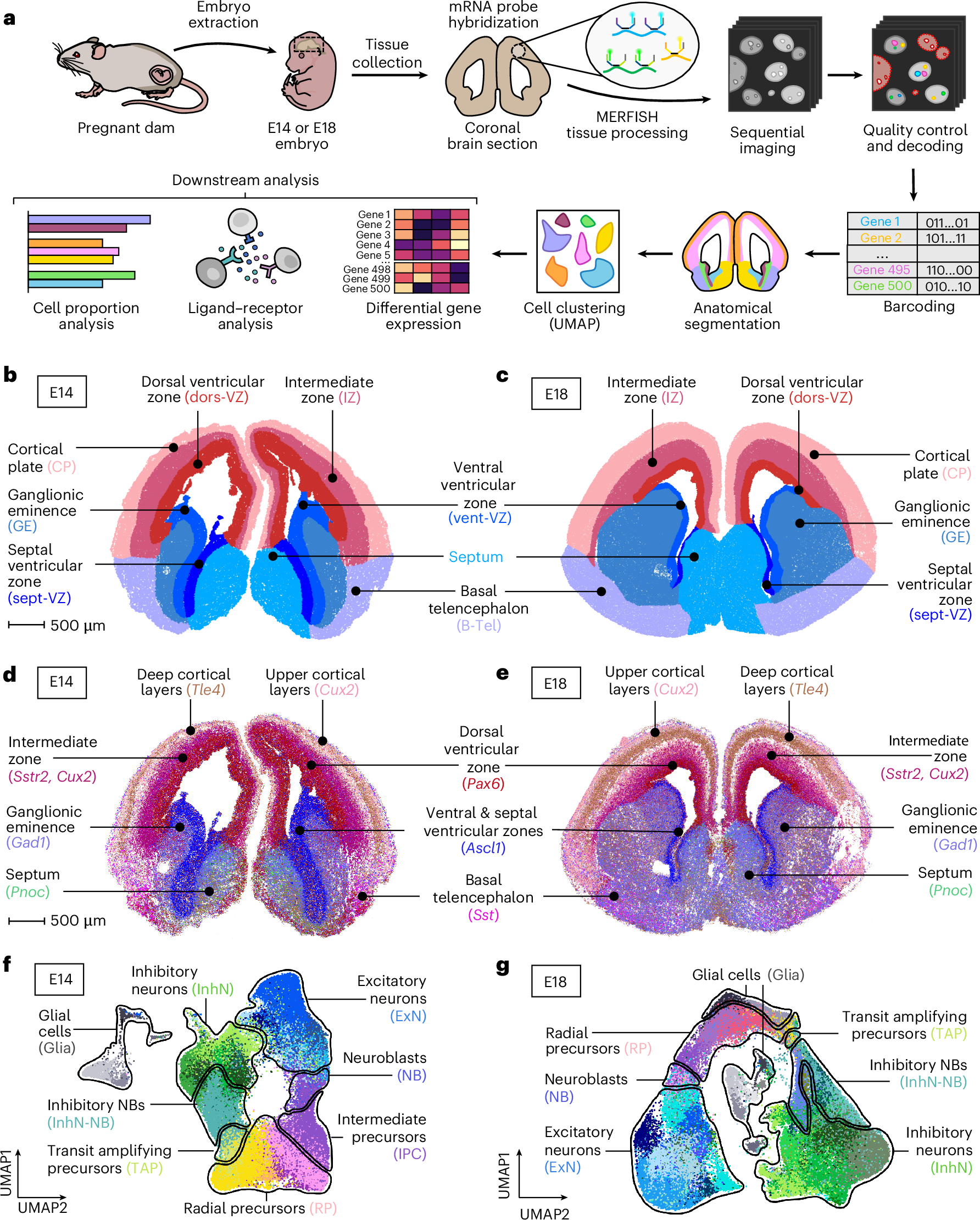
a, Schematic representation of the experimental design. Tissue was collected from E14 and E18 embryos. A total of n = 3 mice at each timepoint were used for MERFISH analysis. b,c, Spatial visualization of the major anatomical regions outlined during sample processing steps in E14 (b) and E18 (c) mice. Scale bar, 500 µm. d,e, Molecular localization of select genes used to identify major anatomical regions in the E14 (d) and E18 (e) brain. Each dot represents a captured gene. Scale bar, 500 µm. f, UMAP representation of broad cell type classes analyzed in the E14 brain, with cell subtypes labeled by color (n = 23 clusters). g, UMAP representation of broad cell type classes analyzed in the E18 brain, with cell subtypes labeled by color (n = 40 clusters).
“Our study establishes a detailed spatial transcriptomic resource of immune gene networks during a critical window of embryonic brain development,” says Kalish. “Unlike previous atlases focused on the adult brain, our dataset captures dynamic immune signaling interactions at a stage when the brain is highly vulnerable.”
The team integrated in situ spatial transcriptomics (MERFISH) with single cell RNA-seq data. This dual-modality approach enabled them to (1) create a developmental cell atlas of immune gene expression in the embryonic mouse brain during mid- and late-gestation; (2) map the spatial location of neurodevelopmental-relevant genes in the developing brain; (3) reveal sex-specific responses of the developing brain to maternal gut-immune disruptions; (4) nominate a specific pathway – known as the CXCL12/CXCR7 signaling pathway – as an important mediator of abnormal neural differentiation.
“As a neonatologist, this work adds to our understanding early-life environmental factors that may impact neurodevelopmental potential and lends insights for potential interventions”, added Kalish.
Source – Boston Children’s Hospital
Availability – These data are accessible to explore via a web interface: https://neurodevelopment.shinyapps.io/DevelopingBrainImmuneAtlas/.


The maternal microbiome and immune system have both independent and synergistic effects on fetal brain health, changes in the mother’s immune system have been linked to an increased risk of neurodevelopmental disorders in children. A new study, published today in Nature Neuroscience, expands our understanding of this “gut-immune axis” by mapping the impact of stressors during pregnancy, namely changes in the microbiome and activation of the immune system, on the neuroimmune landscape of the developing fetal brain.
The research team, from the Division of Newborn Medicine at Boston Children’s Hospital and the SickKids Research Institute, have discovered notable sex-specific responses, including vulnerability for a specific immune pathway in the male brain that could be a potential target for early intervention.
MERFISH of the embryonic mouse brain at E14 and E18
a, Schematic representation of the experimental design. Tissue was collected from E14 and E18 embryos. A total of n = 3 mice at each timepoint were used for MERFISH analysis. b,c, Spatial visualization of the major anatomical regions outlined during sample processing steps in E14 (b) and E18 (c) mice. Scale bar, 500 µm. d,e, Molecular localization of select genes used to identify major anatomical regions in the E14 (d) and E18 (e) brain. Each dot represents a captured gene. Scale bar, 500 µm. f, UMAP representation of broad cell type classes analyzed in the E14 brain, with cell subtypes labeled by color (n = 23 clusters). g, UMAP representation of broad cell type classes analyzed in the E18 brain, with cell subtypes labeled by color (n = 40 clusters).
The team integrated in situ spatial transcriptomics (MERFISH) with single cell RNA-seq data. This dual-modality approach enabled them to (1) create a developmental cell atlas of immune gene expression in the embryonic mouse brain during mid- and late-gestation; (2) map the spatial location of neurodevelopmental-relevant genes in the developing brain; (3) reveal sex-specific responses of the developing brain to maternal gut-immune disruptions; (4) nominate a specific pathway – known as the CXCL12/CXCR7 signaling pathway – as an important mediator of abnormal neural differentiation.
Source – Boston Children’s Hospital
Availability – These data are accessible to explore via a web interface: https://neurodevelopment.shinyapps.io/DevelopingBrainImmuneAtlas/.
Kukreja B, Jeon S, Cao W, Rusu B, Harrison CF, Ghazisaeidi S, Tahmasian N, Feng MY, Chan R, Loan A, Johnston WB, Padhy S, Rakoff-Nahoum S, Wang J, Yim YS, Kalish BT. (2026) Spatial transcriptomics of the developing mouse brain immune landscape reveals effects of maternal immune activation and microbiome depletion. Nature Neuroscience [Epub ahead of print]. [article]
Related Posts
RNAmel simplifies RNA sequencing data analysis with a reproducible, integrated platform
Heart failure reshapes bone marrow to create inflammation-related immune memory
Overlooked DNA regions surrounding ‘microexons’ hold missing pieces of autism’s genetic puzzle
RiboBA improves detection of hidden protein-coding regions using ribosome profiling
An anti-senescence gene may fight spinal disc deterioration
Scientists identify the neuroblastoma cells that signal poor outcomes
Novel biosensor uses DNA ‘barcodes’ to uncover disease signals in cells
CellART extracts single-cell information from high-resolution spatial transcriptomics
Cell2space restores spatial context to single-cell RNA sequencing data
Urine test may improve bladder cancer diagnosis and guide treatment decisions
DeCoRE – a computational method to resolve RNA structural heterogeneity from RNA structure probing data by direct RNA sequencing
Tracing 700 million years of cell evolution
How should biological replicates be handled in long-read RNA sequencing?
RNA-Seq reveals a new way the developing heart controls its growth
The Human RNome Project Consortium seeks to publish first-ever complete sequence of the body’s RNA
scFlowReport brings multiple single-cell RNA sequencing analyses into one workflow
Long-read RNA sequencing reveals how neurons choose alternative RNA endings
DeOPUS improves cell-type estimates from bulk RNA sequencing
ResolVI improves accuracy in spatial transcriptomics analysis
RNA sequencing reveals transcriptional allele differences in prostate cancer stem cells
Stay Connected
Submit a Post to the Blog
Recent Posts
Subscribe to the RNA-Seq Blog
RNA-Seq Products & Services