

Atherosclerosis, the buildup of fatty deposits and inflammation in the arteries, is the main driver of many cardiovascular diseases. These plaques can grow, rupture, and block blood flow, leading to life-threatening conditions like heart attacks and strokes. Understanding which cells are involved in plaque development is critical for finding better ways to prevent and treat these diseases.
Researchers at the Helmholtz Zentrum München have created a detailed single-cell atlas of human atherosclerotic plaques using RNA sequencing. This atlas includes about 250,000 cells with high-quality annotations, providing one of the most comprehensive views of plaque biology to date.
Workflow to construct the single-cell plaque atlas
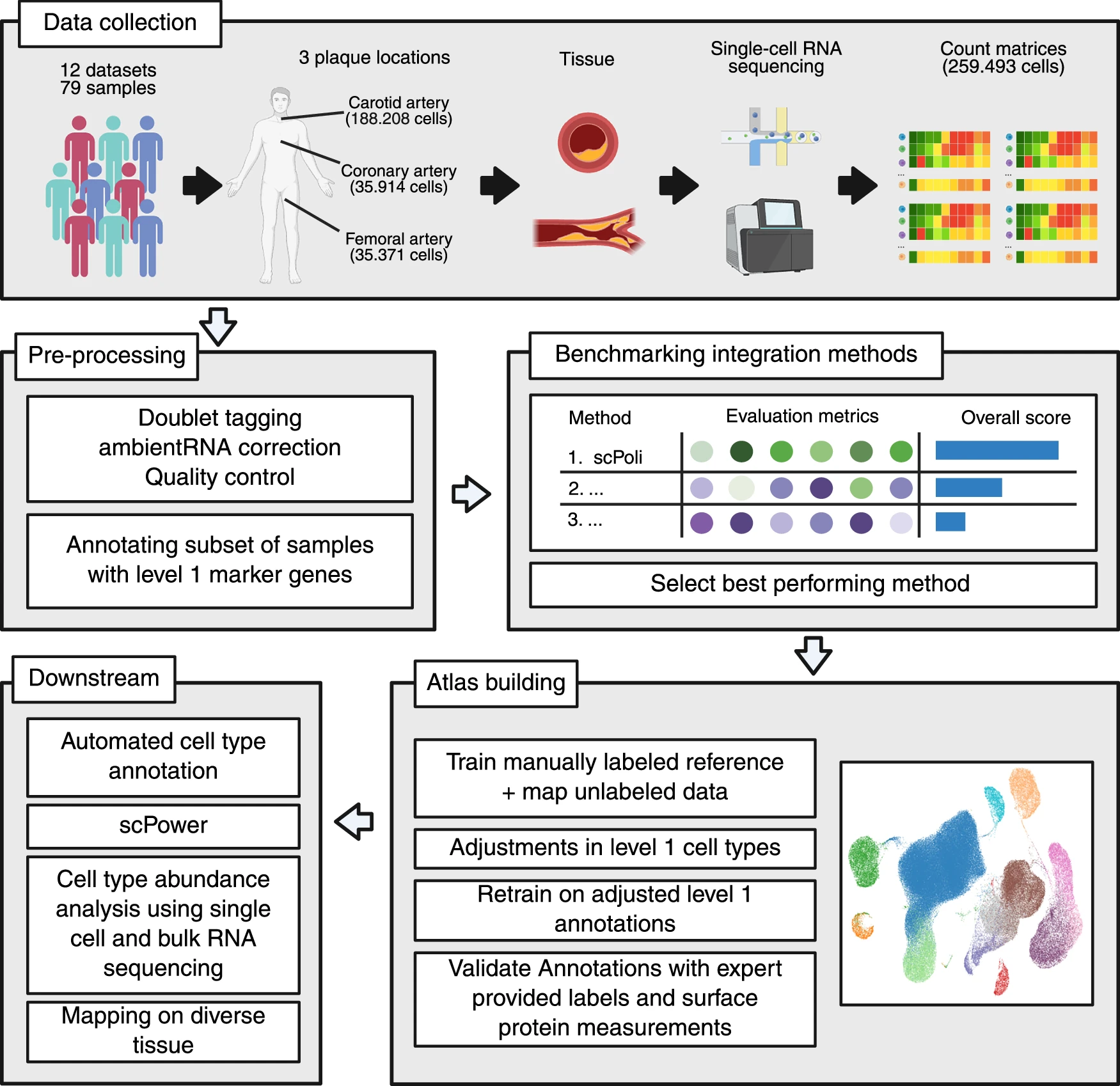
(1) All publicly available scRNA-seq datasets from atherosclerotic plaques from carotid, coronary and femoral arteries were collected from the NCBI GEO database. (2) The same pre-processing pipeline was applied to all datasets. Doublets were tagged, counts were corrected for ambient RNA and cells and genes were filtered as described in the original publication. A subset of samples was manually annotated with level 1 marker genes for the subsequent benchmark of integration methods. (3) A benchmark of integration methods on five batch correction and five biological conservation metrics identified the best performing method, scPoli. (4) ScPoli was applied on the subset of annotated samples to build the atlas. Subsequently, we made adjustments on the level 1 cell types, retrained the reference and mapped the yet unclear cells. The resulting atlas was validated by expert provided labels and surface protein measurements. (5) The annotated plaque atlas was used in the following downstream applications: First, automatic cell type annotation, which was validated with an independent dataset (Bashore et al.) and included in the subsequent reference. This reference was subclustered to define finer-grained level 2 cell types, which were then used for bulk deconvolution and abundance analysis. Lastly, the atlas was used to design future experiments with scPower and mapped on non-lesion arteries and diverse tissue compendium. Created in BioRender. Träuble, K. (2025)
By analyzing this massive dataset, the team identified unique cell types and markers that play important roles in disease progression. For example, they found specialized neutrophils with distinct markers, a cluster of endothelial cells that promote new blood vessel formation in advanced plaques, and subsets of macrophages with specific roles in inflammation. They also discovered that fibromyocytes, a type of structural cell, exist only in vascular tissue.
This resource not only helps scientists better understand the cellular makeup of plaques but also makes it easier to analyze new datasets, design stronger experiments, and interpret bulk RNA sequencing data. To support future research, the team has also provided an interactive web tool so other scientists can explore these findings.
The atlas represents an important step toward uncovering how different cell types drive plaque growth and instability, opening up new possibilities for targeted therapies against cardiovascular disease.
Availability – The fully processed atlas is available through the CELLxGENE portal at https://cellxgene.cziscience.com/collections/db70986c-7d91-49fe-a399-a4730be394ac.
Traeuble K, Munz M, Pauli J, Sachs N, Vafadarnejad E, Carrillo-Roa T, Maegdefessel L, Kastner P, Heinig M. (2025) Integrated single-cell atlas of human atherosclerotic plaques. Nat Commun 16(1): 8255. [article]
Related Posts




Atherosclerosis, the buildup of fatty deposits and inflammation in the arteries, is the main driver of many cardiovascular diseases. These plaques can grow, rupture, and block blood flow, leading to life-threatening conditions like heart attacks and strokes. Understanding which cells are involved in plaque development is critical for finding better ways to prevent and treat these diseases.
Researchers at the Helmholtz Zentrum München have created a detailed single-cell atlas of human atherosclerotic plaques using RNA sequencing. This atlas includes about 250,000 cells with high-quality annotations, providing one of the most comprehensive views of plaque biology to date.
Workflow to construct the single-cell plaque atlas
(1) All publicly available scRNA-seq datasets from atherosclerotic plaques from carotid, coronary and femoral arteries were collected from the NCBI GEO database. (2) The same pre-processing pipeline was applied to all datasets. Doublets were tagged, counts were corrected for ambient RNA and cells and genes were filtered as described in the original publication. A subset of samples was manually annotated with level 1 marker genes for the subsequent benchmark of integration methods. (3) A benchmark of integration methods on five batch correction and five biological conservation metrics identified the best performing method, scPoli. (4) ScPoli was applied on the subset of annotated samples to build the atlas. Subsequently, we made adjustments on the level 1 cell types, retrained the reference and mapped the yet unclear cells. The resulting atlas was validated by expert provided labels and surface protein measurements. (5) The annotated plaque atlas was used in the following downstream applications: First, automatic cell type annotation, which was validated with an independent dataset (Bashore et al.) and included in the subsequent reference. This reference was subclustered to define finer-grained level 2 cell types, which were then used for bulk deconvolution and abundance analysis. Lastly, the atlas was used to design future experiments with scPower and mapped on non-lesion arteries and diverse tissue compendium. Created in BioRender. Träuble, K. (2025)
By analyzing this massive dataset, the team identified unique cell types and markers that play important roles in disease progression. For example, they found specialized neutrophils with distinct markers, a cluster of endothelial cells that promote new blood vessel formation in advanced plaques, and subsets of macrophages with specific roles in inflammation. They also discovered that fibromyocytes, a type of structural cell, exist only in vascular tissue.
This resource not only helps scientists better understand the cellular makeup of plaques but also makes it easier to analyze new datasets, design stronger experiments, and interpret bulk RNA sequencing data. To support future research, the team has also provided an interactive web tool so other scientists can explore these findings.
The atlas represents an important step toward uncovering how different cell types drive plaque growth and instability, opening up new possibilities for targeted therapies against cardiovascular disease.
Availability – The fully processed atlas is available through the CELLxGENE portal at https://cellxgene.cziscience.com/collections/db70986c-7d91-49fe-a399-a4730be394ac.
Traeuble K, Munz M, Pauli J, Sachs N, Vafadarnejad E, Carrillo-Roa T, Maegdefessel L, Kastner P, Heinig M. (2025) Integrated single-cell atlas of human atherosclerotic plaques. Nat Commun 16(1): 8255. [article]
Related Posts
Recent Posts
 RNAmel simplifies RNA sequencing data analysis with a reproducible, integrated platform
 Heart failure reshapes bone marrow to create inflammation-related immune memory
 Overlooked DNA regions surrounding ‘microexons’ hold missing pieces of autism’s genetic puzzle
 RiboBA improves detection of hidden protein-coding regions using ribosome profiling
 An anti-senescence gene may fight spinal disc deterioration
 Bulk RNA-seq Analysis: A Complete Beginner’s Guide
RNA-Seq Products & Services



Stay Connected