

Understanding how DNA is regulated inside individual cells is essential for decoding the complexity of tissues and diseases. One major part of that regulation involves chromatin, the structure that packages DNA and controls which genes are turned on or off. However, studying chromatin in single cells has been challenging, especially when trying to track how proteins interact with DNA. Traditional methods often require expensive equipment, lengthy protocols, or don’t work well with rare chromatin features.
Now, a team of scientists led researchers at the University of Hong Kong has introduced a breakthrough method that makes this process more efficient, scalable, and accessible. The technique is called indexed tagmentation-based single-cell CUT&Tag-sequencing, or IT-scC&T-seq for short.
What Is IT-scC&T-seq and Why Is It Important?
IT-scC&T-seq allows researchers to profile protein-DNA interactions, such as those involving histone modifications and transcription factors, at single-cell resolution. These proteins help determine how tightly DNA is wound up, which in turn influences which genes are active. Unlike earlier methods that required high-cost instruments or specialized microfluidics, IT-scC&T-seq is plate-based and uses a clever system of three rounds of barcoding. This allows many samples to be processed together in one batch while still keeping track of which cell each dataset came from.
Design and benchmarking of IT-scC&T-seq
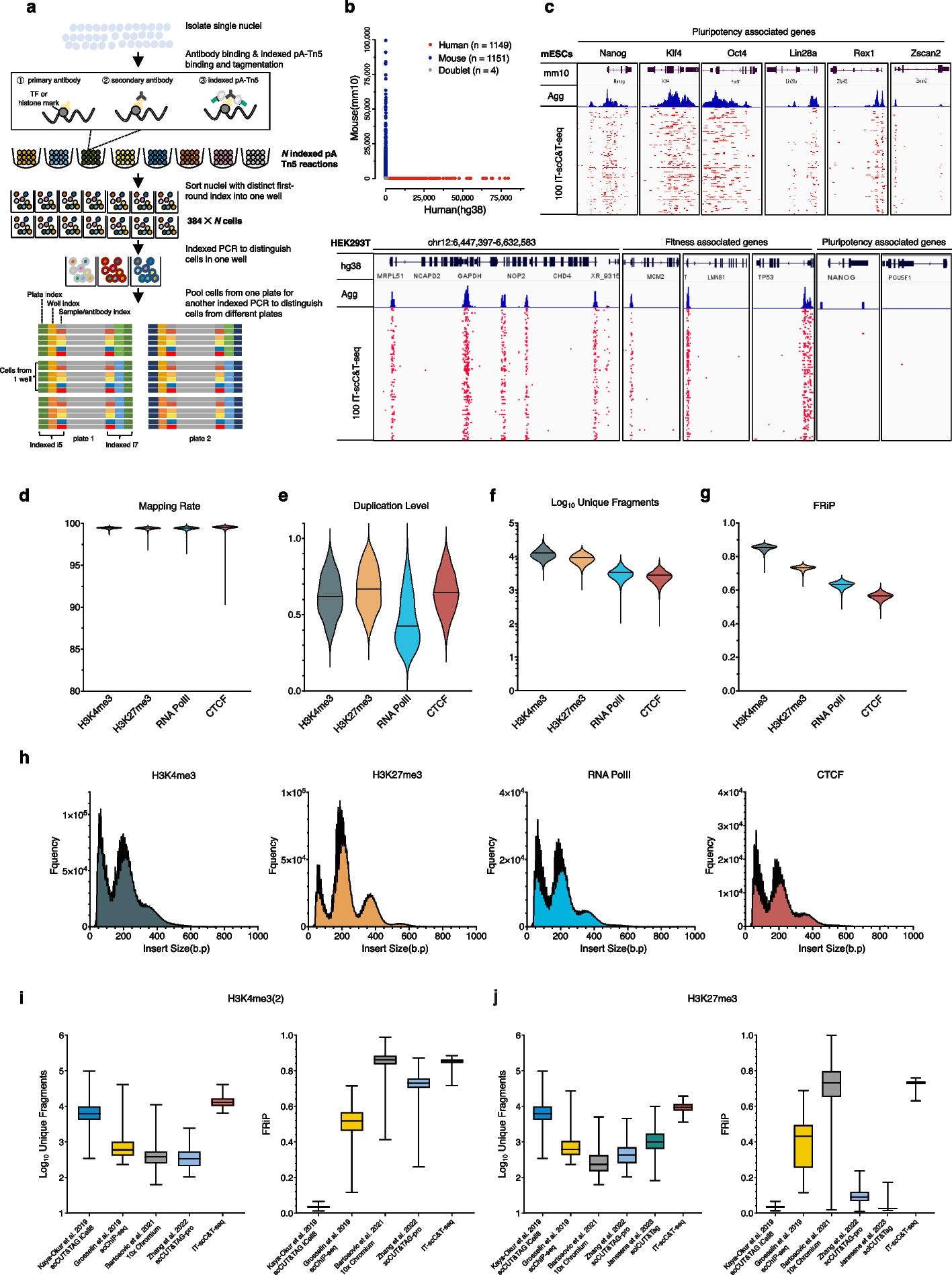
a Overview of the IT-scC&T-seq workflow. Isolated nuclei are incubated with primary and secondary antibodies, then split into multiple tagmentation reactions (number of reactions = N) using uniquely barcoded pA-Tn5 complexes. Transposed nuclei from each tagmentation reaction are individually and sequentially distributed into 384-well plates via fluorescence-activated nuclei sorting. After lysis, a first round of barcoded PCR is performed to label wells, followed by pooling and a second round of PCR to add Illumina adapters. Libraries are then sequenced. b Species mixing experiment using H3K4me3-targeted IT-scC&T-seq on a 1:1 mixture of HEK293T and mESCs. The scatter plot shows the number of reads per cell aligned to human vs mouse genomes. 99.83% of cells were species-specific, with 1149 humans, 1151 mouse, and 4 unassigned cells. c Genome browser tracks displaying single-cell and aggregated profiles. Pluripotency genes (e.g., Nanog, Klf4, Oct4, Lin28, Rex1, Zscan2) were enriched in mESCs. Housekeeping genes (e.g., GAPDH, MCM2, LMNB1, TP53) were enriched in HEK293T. Per cell quality control metrics of IT-scC&T-seq in K562 cells across histone marks (H3K4me3, H3K27me3) and transcription factors (RNA Pol II, CTCF): mapping rate (d), duplication rate (e), log10 of unique fragments (f), and FRiP scores (g). h Fragment length distributions demonstrating nucleosomal periodicity across histone marks and TFs. Comparisons of library complexity and FRiP between IT-scC&T-seq and other single-cell chromatin profiling methods for H3K4me3 or H3K4me2 (i) and H3K27me3 (j)
Key Advantages of the Method
One standout feature of this method is its ability to identify lamina-associated domains (LADs); sections of the genome that are typically silenced and tucked away near the nuclear envelope. These domains are usually present in low abundance and have been very difficult to detect in single cells. With IT-scC&T-seq, scientists can now study LADs alongside more active chromatin marks in the same cells.
Another strength of IT-scC&T-seq is its high specificity and throughput, which makes it ideal for analyzing tissues that contain many different cell types. The team demonstrated this by applying the method to the adult mouse mammary gland, a tissue that includes stem cells, progenitor cells, and differentiated cells. They found that each cell type has a unique chromatin landscape, and they were even able to track lineage-regulatory changes—in other words, changes that help determine a cell’s identity and future path.
Expanding Access to High-Resolution Chromatin Research
By combining protein-DNA interaction profiling with RNA sequencing or other single-cell techniques, IT-scC&T-seq could help scientists build a complete picture of how gene regulation changes across cell types, developmental stages, or disease states. This is especially relevant for studying cancer, developmental biology, and regenerative medicine, where understanding the regulation of individual cells is key.
Most importantly, the method is cost-effective and doesn’t rely on hard-to-access equipment, meaning it could be adopted more widely in both academic and clinical research settings.
Ma J, Jin W, Rong L, Gao Z, Hazrat Z, Shakhawat HM, Long F, Zhang Z, Huang J, Lu X, Jin G, Zhou Z. (2025) IT-scC&T-seq streamlines scalable, parallel profiling of protein-DNA interactions in single cells. Genome Biol 26(1):196. [article]
Related Posts






Understanding how DNA is regulated inside individual cells is essential for decoding the complexity of tissues and diseases. One major part of that regulation involves chromatin, the structure that packages DNA and controls which genes are turned on or off. However, studying chromatin in single cells has been challenging, especially when trying to track how proteins interact with DNA. Traditional methods often require expensive equipment, lengthy protocols, or don’t work well with rare chromatin features.
Now, a team of scientists led researchers at the University of Hong Kong has introduced a breakthrough method that makes this process more efficient, scalable, and accessible. The technique is called indexed tagmentation-based single-cell CUT&Tag-sequencing, or IT-scC&T-seq for short.
What Is IT-scC&T-seq and Why Is It Important?
IT-scC&T-seq allows researchers to profile protein-DNA interactions, such as those involving histone modifications and transcription factors, at single-cell resolution. These proteins help determine how tightly DNA is wound up, which in turn influences which genes are active. Unlike earlier methods that required high-cost instruments or specialized microfluidics, IT-scC&T-seq is plate-based and uses a clever system of three rounds of barcoding. This allows many samples to be processed together in one batch while still keeping track of which cell each dataset came from.
Design and benchmarking of IT-scC&T-seq
a Overview of the IT-scC&T-seq workflow. Isolated nuclei are incubated with primary and secondary antibodies, then split into multiple tagmentation reactions (number of reactions = N) using uniquely barcoded pA-Tn5 complexes. Transposed nuclei from each tagmentation reaction are individually and sequentially distributed into 384-well plates via fluorescence-activated nuclei sorting. After lysis, a first round of barcoded PCR is performed to label wells, followed by pooling and a second round of PCR to add Illumina adapters. Libraries are then sequenced. b Species mixing experiment using H3K4me3-targeted IT-scC&T-seq on a 1:1 mixture of HEK293T and mESCs. The scatter plot shows the number of reads per cell aligned to human vs mouse genomes. 99.83% of cells were species-specific, with 1149 humans, 1151 mouse, and 4 unassigned cells. c Genome browser tracks displaying single-cell and aggregated profiles. Pluripotency genes (e.g., Nanog, Klf4, Oct4, Lin28, Rex1, Zscan2) were enriched in mESCs. Housekeeping genes (e.g., GAPDH, MCM2, LMNB1, TP53) were enriched in HEK293T. Per cell quality control metrics of IT-scC&T-seq in K562 cells across histone marks (H3K4me3, H3K27me3) and transcription factors (RNA Pol II, CTCF): mapping rate (d), duplication rate (e), log10 of unique fragments (f), and FRiP scores (g). h Fragment length distributions demonstrating nucleosomal periodicity across histone marks and TFs. Comparisons of library complexity and FRiP between IT-scC&T-seq and other single-cell chromatin profiling methods for H3K4me3 or H3K4me2 (i) and H3K27me3 (j)
Key Advantages of the Method
One standout feature of this method is its ability to identify lamina-associated domains (LADs); sections of the genome that are typically silenced and tucked away near the nuclear envelope. These domains are usually present in low abundance and have been very difficult to detect in single cells. With IT-scC&T-seq, scientists can now study LADs alongside more active chromatin marks in the same cells.
Another strength of IT-scC&T-seq is its high specificity and throughput, which makes it ideal for analyzing tissues that contain many different cell types. The team demonstrated this by applying the method to the adult mouse mammary gland, a tissue that includes stem cells, progenitor cells, and differentiated cells. They found that each cell type has a unique chromatin landscape, and they were even able to track lineage-regulatory changes—in other words, changes that help determine a cell’s identity and future path.
Expanding Access to High-Resolution Chromatin Research
By combining protein-DNA interaction profiling with RNA sequencing or other single-cell techniques, IT-scC&T-seq could help scientists build a complete picture of how gene regulation changes across cell types, developmental stages, or disease states. This is especially relevant for studying cancer, developmental biology, and regenerative medicine, where understanding the regulation of individual cells is key.
Most importantly, the method is cost-effective and doesn’t rely on hard-to-access equipment, meaning it could be adopted more widely in both academic and clinical research settings.
Ma J, Jin W, Rong L, Gao Z, Hazrat Z, Shakhawat HM, Long F, Zhang Z, Huang J, Lu X, Jin G, Zhou Z. (2025) IT-scC&T-seq streamlines scalable, parallel profiling of protein-DNA interactions in single cells. Genome Biol 26(1):196. [article]
Related Posts
Recent Posts
 RNAmel simplifies RNA sequencing data analysis with a reproducible, integrated platform
 Heart failure reshapes bone marrow to create inflammation-related immune memory
 Overlooked DNA regions surrounding ‘microexons’ hold missing pieces of autism’s genetic puzzle
 RiboBA improves detection of hidden protein-coding regions using ribosome profiling
 An anti-senescence gene may fight spinal disc deterioration
 Bulk RNA-seq Analysis: A Complete Beginner’s Guide
RNA-Seq Products & Services



Stay Connected