

Cells do not translate all messenger RNAs at the same speed. The rate at which ribosomes move along an mRNA can change depending on the surrounding sequence, the availability of tRNAs, the growing protein chain, or external stresses like antibiotics. These pauses and slowdowns matter because they influence how much protein is made and how well it folds.
Researchers at the Institut National de la Santé et de la Recherche Médicale, France have developed and refined a method called inverse toeprinting coupled to next-generation sequencing, known as iTP-seq, to study these effects in bacteria. The approach is designed to complement ribosome profiling, which measures translation inside living cells, by providing a powerful in vitro way to map where ribosomes pause during protein synthesis.
Overview of the iTP-seq workflow to study context-dependent translational events in vitro
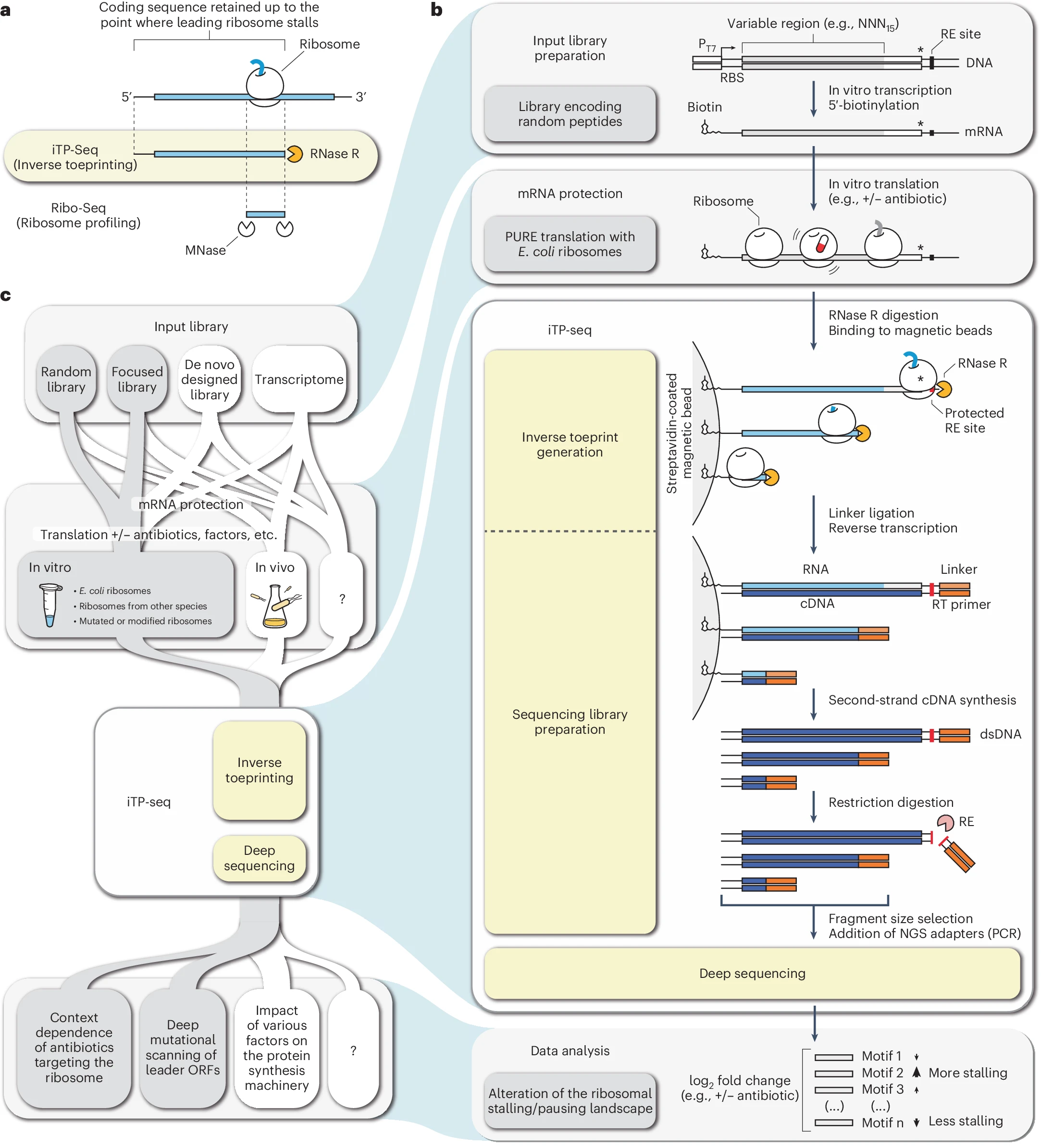
a, Basic principle of iTP-seq, whereby a highly processive RNA exonuclease (RNase R) is used to locate the leading ribosomes on mRNAs with codon resolution by degrading transcripts from the 3′ end. In contrast with Ribo-seq, in which short mRNA footprints generated by an RNA endonuclease are mapped to a sequenced genome, the entire coding region up to the leading ribosome is preserved and sequenced during iTP-seq. b, Exploratory iTP-seq workflow for identifying the context dependence of a ribosome-targeting antibiotic, as described in our previous study on the aromatic polyketide antibiotic TcmX. The input library encodes peptides with a 15-residue long random region (NNN15), and in vitro translation is carried out by using a commercial PURExpress system. The invariant iTP-seq step is highlighted in yellow. RNase R cleaves the mRNA 17–19 nt downstream of the ribosomal P site. Ribosomes that have reached the stop codon (asterisk) protect a restriction site (restriction enzyme (RE) site, shown in red) from RNase R digestion, ensuring that unwanted fragments corresponding to translation termination complexes are removed upon treating cDNAs with the corresponding restriction enzyme. Data analysis relies on the comparison between drug-treated and untreated samples to identify nascent amino acid motifs that cause ribosomes to stall in the presence of the antibiotic. Arrows in the data-analysis panel indicate greater (up arrow) or lower (down arrow) relative stalling for a given motif. Arrow thickness is proportional to the change in frequency of a given motif in the treated versus untreated samples. c, Possible workflows incorporating iTP-seq in combination with various input libraries, mRNA-protection approaches (e.g., translation) and data-analysis pipelines. Workflows tested in our laboratory are shaded in gray, and additional, untested workflows are shown in white. The iTP-seq step itself is highlighted in yellow. dsDNA, double-stranded DNA; MNase, micrococcal nuclease; RT, reverse transcription.
In iTP-seq, bacterial mRNAs are translated in a test tube. Ribosomes protect the portion of the RNA they are translating, while an enzyme called RNase R digests the rest of the RNA from the 3′ end. What remains are ribosome-protected fragments, called inverse toeprints, that mark the exact position of the leading ribosome on each message. These fragments are then analyzed by RNA sequencing, allowing researchers to pinpoint ribosome positions at single-codon resolution.
A key advantage of iTP-seq is that it does not require prior knowledge of the RNA sequences being translated. This means scientists can use fully custom-designed libraries rather than relying on existing genomes. As a result, the method is especially useful for testing how specific sequence contexts or antibiotics affect translation.
The authors present a detailed, standardized protocol that covers library preparation, in vitro translation, fragment generation, sequencing, and data analysis. They also show how iTP-seq can be integrated into broader workflows to study context-dependent translation inhibitors, such as antibiotics that stall ribosomes at specific sequences. Together, this makes iTP-seq a flexible and scalable tool for exploring how bacterial translation is regulated at high resolution.
Availability – Code used for iTP-seq analysis is published as a python library (https://pypi.org/project/itpseq) and is available for reuse under the GPLv3 license.
Gillard M, Renault TT, Innis CA. (2026) iTP-seq a scalable profiling workflow to characterize bacterial translation landscapes in vitro. Nature Protocols [Epub ahead of print]. [article]
Related Posts




Cells do not translate all messenger RNAs at the same speed. The rate at which ribosomes move along an mRNA can change depending on the surrounding sequence, the availability of tRNAs, the growing protein chain, or external stresses like antibiotics. These pauses and slowdowns matter because they influence how much protein is made and how well it folds.
Researchers at the Institut National de la Santé et de la Recherche Médicale, France have developed and refined a method called inverse toeprinting coupled to next-generation sequencing, known as iTP-seq, to study these effects in bacteria. The approach is designed to complement ribosome profiling, which measures translation inside living cells, by providing a powerful in vitro way to map where ribosomes pause during protein synthesis.
Overview of the iTP-seq workflow to study context-dependent translational events in vitro
a, Basic principle of iTP-seq, whereby a highly processive RNA exonuclease (RNase R) is used to locate the leading ribosomes on mRNAs with codon resolution by degrading transcripts from the 3′ end. In contrast with Ribo-seq, in which short mRNA footprints generated by an RNA endonuclease are mapped to a sequenced genome, the entire coding region up to the leading ribosome is preserved and sequenced during iTP-seq. b, Exploratory iTP-seq workflow for identifying the context dependence of a ribosome-targeting antibiotic, as described in our previous study on the aromatic polyketide antibiotic TcmX. The input library encodes peptides with a 15-residue long random region (NNN15), and in vitro translation is carried out by using a commercial PURExpress system. The invariant iTP-seq step is highlighted in yellow. RNase R cleaves the mRNA 17–19 nt downstream of the ribosomal P site. Ribosomes that have reached the stop codon (asterisk) protect a restriction site (restriction enzyme (RE) site, shown in red) from RNase R digestion, ensuring that unwanted fragments corresponding to translation termination complexes are removed upon treating cDNAs with the corresponding restriction enzyme. Data analysis relies on the comparison between drug-treated and untreated samples to identify nascent amino acid motifs that cause ribosomes to stall in the presence of the antibiotic. Arrows in the data-analysis panel indicate greater (up arrow) or lower (down arrow) relative stalling for a given motif. Arrow thickness is proportional to the change in frequency of a given motif in the treated versus untreated samples. c, Possible workflows incorporating iTP-seq in combination with various input libraries, mRNA-protection approaches (e.g., translation) and data-analysis pipelines. Workflows tested in our laboratory are shaded in gray, and additional, untested workflows are shown in white. The iTP-seq step itself is highlighted in yellow. dsDNA, double-stranded DNA; MNase, micrococcal nuclease; RT, reverse transcription.
In iTP-seq, bacterial mRNAs are translated in a test tube. Ribosomes protect the portion of the RNA they are translating, while an enzyme called RNase R digests the rest of the RNA from the 3′ end. What remains are ribosome-protected fragments, called inverse toeprints, that mark the exact position of the leading ribosome on each message. These fragments are then analyzed by RNA sequencing, allowing researchers to pinpoint ribosome positions at single-codon resolution.
A key advantage of iTP-seq is that it does not require prior knowledge of the RNA sequences being translated. This means scientists can use fully custom-designed libraries rather than relying on existing genomes. As a result, the method is especially useful for testing how specific sequence contexts or antibiotics affect translation.
The authors present a detailed, standardized protocol that covers library preparation, in vitro translation, fragment generation, sequencing, and data analysis. They also show how iTP-seq can be integrated into broader workflows to study context-dependent translation inhibitors, such as antibiotics that stall ribosomes at specific sequences. Together, this makes iTP-seq a flexible and scalable tool for exploring how bacterial translation is regulated at high resolution.
Availability – Code used for iTP-seq analysis is published as a python library (https://pypi.org/project/itpseq) and is available for reuse under the GPLv3 license.
Gillard M, Renault TT, Innis CA. (2026) iTP-seq a scalable profiling workflow to characterize bacterial translation landscapes in vitro. Nature Protocols [Epub ahead of print]. [article]
Related Posts
Recent Posts
 Machine learning reveals how RNA splicing is regulated in individual cell types
 Illumina releases SpliceAI2 to help advance rare disease research
 Postdoctoral Fellow – Long-Read RNA Methods Development for Neurodegeneration
 Novel biosensor uses DNA ‘barcodes’ to uncover disease signals in cells
 GP-DHT uses single-cell RNA sequencing to map gene regulatory networks
 Boehringer taps Envisagenics’ AI to mine RNA splicing for cancer targets
RNA-Seq Products & Services


Stay Connected