Largest study on brain aging points to possible connections between diet, inflammation, and brain health
Scientists at the Allen Institute have identified specific cell types in the brain of mice that undergo major changes as they age, along with a specific hot spot where many of those changes occur. The discoveries, published in the journal Nature, could pave the way for future therapies to slow or manage the aging process in the brain.
The scientists discovered dozens of specific cell types, mostly glial cells, known as brain support cells, that underwent significant gene expression changes with age. Those strongly affected included microglia and border-associated macrophages, oligodendrocytes, tanycytes, and ependymal cells.
They found that in aging brains, genes associated with inflammation increased in activity while those related to neuronal structure and function decreased.
They also discovered a specific hot spot combining both the decrease in neuronal function and the increase in inflammation in the hypothalamus. The most significant gene expression changes were found in cell types near the third ventricle of the hypothalamus, including tanycytes, ependymal cells, and neurons known for their role in food intake, energy homeostasis, metabolism, and how our bodies use nutrients.
Transcriptomic cell types in young adult and aged mouse brains

a, Schematic of dissected brain regions profiled in this study, coloured by major brain structure. b, Schematic of library generation and cell annotation workflow. c, Diagram of cell annotation levels based on the ABC-WMB atlas. Bold text indicates the highlighted cell type that is expanded in the subsequent, finer annotation level. d, Schematic of general analysis strategy for identifying cell types vulnerable to ageing. e, UMAP representation of all single-cell transcriptomes included in this study, coloured by cell class, major brain structure and age. f, Summary of the number of all age-DE genes identified from each subclass (for neurons) and supertype (for non-neuronal cells). Bar graphs below age-DE gene counts represent breakdown of each group by major brain structure, age and sex. g, Relationship between the number of age-DE genes and Augur AUC score for all cell types represented in f. Linear model with 95% confidence interval of fit is shown by the light grey shading. h, Histogram of the number of cell types (subclasses for neurons; supertypes for non-neuronal cells) an age-DE gene is significant for. PL + ILA + ORB, prelimbic area + infralimbic area + orbital area; AI, agranular insular area; ACA, anterior cingulate area; RSP, retrosplenial area; HIP-CA, hippocampus; PAR + POST + PRE + ProS + SUB, parasubiculum + postsubiculum + presubiculum + prosubiculum + subiculum; ENT, lateral and medial entorhinal areas; HY, hypothalamus; STRd, dorsal striatum; STRv, ventral striatum; PAL, pallidum; sAMY, striatum-like amygdalar nuclei; PAG + RAmb, periaqueductal grey + midbrain raphe nuclei; SNr + SNc + VTA, substantia nigra, reticular part + substantia nigra, compact part + ventral tegmental area.
This points to a possible connection between diet, lifestyle factors, brain aging, and changes that can influence our susceptibility to age-related brain disorders.
“Our hypothesis is that those cell types are getting less efficient at integrating signals from our environment or from things that we’re consuming,” said Kelly Jin, Ph.D., a scientist at the Allen Institute for Brain Science and lead author of the study. “And that loss of efficiency somehow contributes to what we know as aging in the rest of our body. I think that’s pretty amazing, and I think it’s remarkable that we’re able to find those very specific changes with the methods that we’re using.”
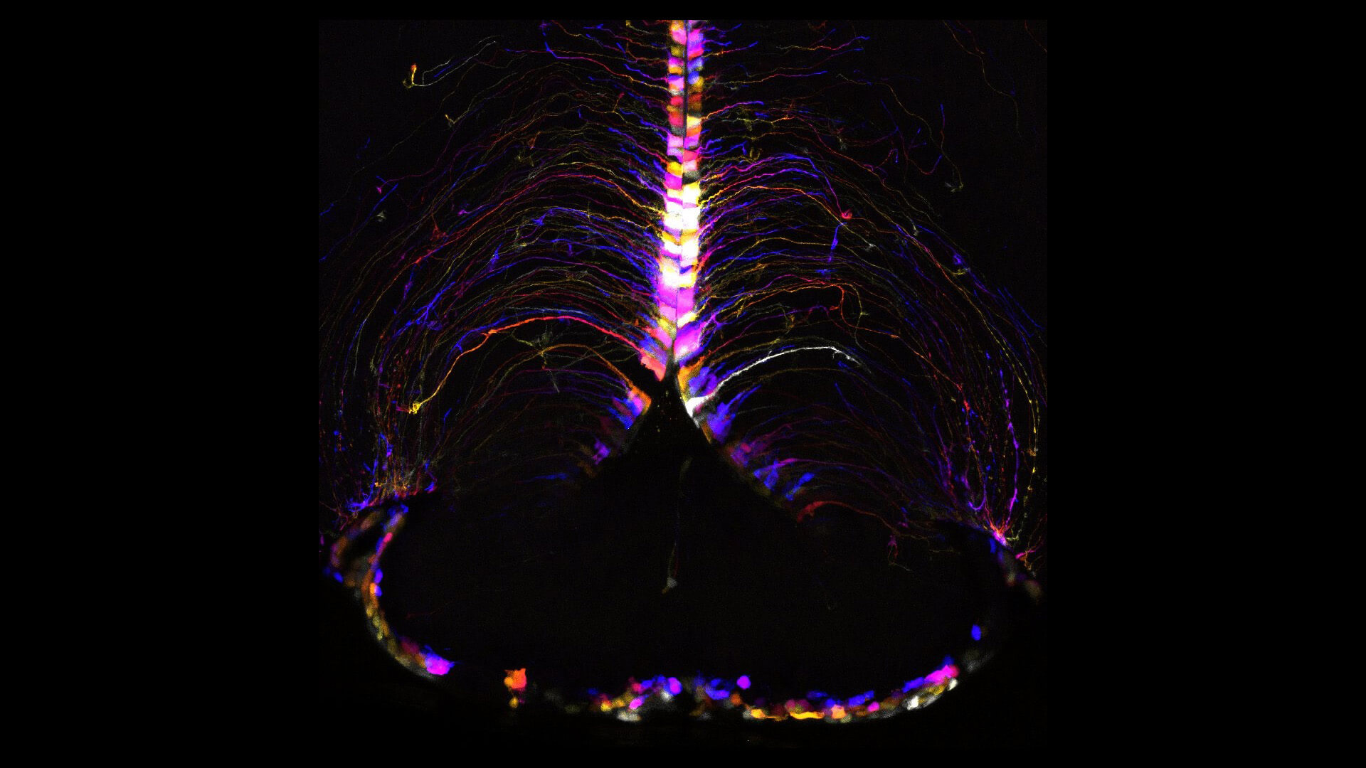
To conduct the study, researchers used cutting-edge single-cell RNA sequencing and advanced brain-mapping tools developed through NIH’s BRAIN Initiative to map over 1.2 million brain cells from young (2 months old) and aged (18 months old) mice across 16 broad brain regions. The aged mice are what scientists consider to be the equivalent of a late middle-aged human. Mouse brains share many similarities with human brains in terms of structure, function, genes, and cell types.
“Aging is the most important risk factor for Alzheimer’s disease and many other devastating brain disorders. These results provide a highly detailed map for which brain cells may be most affected by aging,” said Richard J. Hodes, M.D., director of NIH’s National Institute on Aging. “This new map may fundamentally alter the way scientists think about how aging affects the brain and also provides a guide for developing new treatments for aging-related brain diseases.”
A path toward new therapies
Understanding this hot spot in the hypothalamus makes it a focal point for future study. Along with knowing which cells to specifically target, this could lead to the development of age-related therapeutics, helping to preserve function and prevent neurodegenerative disease.
“We want to develop tools that can target those cell types,” said Hongkui Zeng, Ph.D., executive vice president and director of the Allen Institute for Brain Science. “If we improve the function of those cells, will we be able to delay the aging process?”
The latest findings also align with past studies that link aging to metabolic changes as well as research suggesting that intermittent fasting, balanced diet, or calorie restriction can influence or perhaps increase lifespan.
“It’s not something we directly tested in this study,” said Jin. “But to me, it points to the potential players involved in the process, which I think is a huge deal because this is a very specific, rare population of neurons that express very specific genes that people can develop tools to target and further study.”
Future brain aging research
This study lays the groundwork for new strategies in diet and therapeutic approaches aimed at maintaining brain health into old age, along with more research on the complexities of advanced aging in the brain. As scientists further explore these connections, research may unlock more specific dietary or drug interventions to combat or slow aging on a cellular level.
“The important thing about our study is that we found the key players—the real key players—and the biological substrates for this process,” said Zeng. “Putting the pieces of this puzzle together, you have to find the right players. It’s a beautiful example of why you need to study the brain and the body at this kind of cell type-specific level. Otherwise, changes happening in specific cell types could be averaged out and undetected if you mix different types of cells together.”
Source – The Allen Institute


Largest study on brain aging points to possible connections between diet, inflammation, and brain health
Scientists at the Allen Institute have identified specific cell types in the brain of mice that undergo major changes as they age, along with a specific hot spot where many of those changes occur. The discoveries, published in the journal Nature, could pave the way for future therapies to slow or manage the aging process in the brain.
The scientists discovered dozens of specific cell types, mostly glial cells, known as brain support cells, that underwent significant gene expression changes with age. Those strongly affected included microglia and border-associated macrophages, oligodendrocytes, tanycytes, and ependymal cells.
They found that in aging brains, genes associated with inflammation increased in activity while those related to neuronal structure and function decreased.
They also discovered a specific hot spot combining both the decrease in neuronal function and the increase in inflammation in the hypothalamus. The most significant gene expression changes were found in cell types near the third ventricle of the hypothalamus, including tanycytes, ependymal cells, and neurons known for their role in food intake, energy homeostasis, metabolism, and how our bodies use nutrients.
Transcriptomic cell types in young adult and aged mouse brains
a, Schematic of dissected brain regions profiled in this study, coloured by major brain structure. b, Schematic of library generation and cell annotation workflow. c, Diagram of cell annotation levels based on the ABC-WMB atlas. Bold text indicates the highlighted cell type that is expanded in the subsequent, finer annotation level. d, Schematic of general analysis strategy for identifying cell types vulnerable to ageing. e, UMAP representation of all single-cell transcriptomes included in this study, coloured by cell class, major brain structure and age. f, Summary of the number of all age-DE genes identified from each subclass (for neurons) and supertype (for non-neuronal cells). Bar graphs below age-DE gene counts represent breakdown of each group by major brain structure, age and sex. g, Relationship between the number of age-DE genes and Augur AUC score for all cell types represented in f. Linear model with 95% confidence interval of fit is shown by the light grey shading. h, Histogram of the number of cell types (subclasses for neurons; supertypes for non-neuronal cells) an age-DE gene is significant for. PL + ILA + ORB, prelimbic area + infralimbic area + orbital area; AI, agranular insular area; ACA, anterior cingulate area; RSP, retrosplenial area; HIP-CA, hippocampus; PAR + POST + PRE + ProS + SUB, parasubiculum + postsubiculum + presubiculum + prosubiculum + subiculum; ENT, lateral and medial entorhinal areas; HY, hypothalamus; STRd, dorsal striatum; STRv, ventral striatum; PAL, pallidum; sAMY, striatum-like amygdalar nuclei; PAG + RAmb, periaqueductal grey + midbrain raphe nuclei; SNr + SNc + VTA, substantia nigra, reticular part + substantia nigra, compact part + ventral tegmental area.
This points to a possible connection between diet, lifestyle factors, brain aging, and changes that can influence our susceptibility to age-related brain disorders.
To conduct the study, researchers used cutting-edge single-cell RNA sequencing and advanced brain-mapping tools developed through NIH’s BRAIN Initiative to map over 1.2 million brain cells from young (2 months old) and aged (18 months old) mice across 16 broad brain regions. The aged mice are what scientists consider to be the equivalent of a late middle-aged human. Mouse brains share many similarities with human brains in terms of structure, function, genes, and cell types.
A path toward new therapies
Understanding this hot spot in the hypothalamus makes it a focal point for future study. Along with knowing which cells to specifically target, this could lead to the development of age-related therapeutics, helping to preserve function and prevent neurodegenerative disease.
The latest findings also align with past studies that link aging to metabolic changes as well as research suggesting that intermittent fasting, balanced diet, or calorie restriction can influence or perhaps increase lifespan.
Future brain aging research
This study lays the groundwork for new strategies in diet and therapeutic approaches aimed at maintaining brain health into old age, along with more research on the complexities of advanced aging in the brain. As scientists further explore these connections, research may unlock more specific dietary or drug interventions to combat or slow aging on a cellular level.
Source – The Allen Institute
Jin K, Yao Z, van Velthoven CTJ, Kaplan ES, Glattfelder K, Barlow ST, Boyer G, Carey D, Casper T, Chakka AB, Chakrabarty R, Clark M, Departee M, Desierto M, Gary A, Gloe J, Goldy J, Guilford N, Guzman J, Hirschstein D, Lee C, Liang E, Pham T, Reding M, Ronellenfitch K, Ruiz A, Sevigny J, Shapovalova N, Shulga L, Sulc J, Torkelson A, Tung H, Levi B, Sunkin SM, Dee N, Esposito L, Smith KA, Tasic B, Zeng H. (2025) Brain-wide cell-type-specific transcriptomic signatures of healthy ageing in mice. Nature 638(8049):182-196. [article]
Related Posts
RNAmel simplifies RNA sequencing data analysis with a reproducible, integrated platform
Heart failure reshapes bone marrow to create inflammation-related immune memory
Overlooked DNA regions surrounding ‘microexons’ hold missing pieces of autism’s genetic puzzle
RiboBA improves detection of hidden protein-coding regions using ribosome profiling
An anti-senescence gene may fight spinal disc deterioration
Scientists identify the neuroblastoma cells that signal poor outcomes
Novel biosensor uses DNA ‘barcodes’ to uncover disease signals in cells
CellART extracts single-cell information from high-resolution spatial transcriptomics
Cell2space restores spatial context to single-cell RNA sequencing data
Urine test may improve bladder cancer diagnosis and guide treatment decisions
DeCoRE – a computational method to resolve RNA structural heterogeneity from RNA structure probing data by direct RNA sequencing
Tracing 700 million years of cell evolution
How should biological replicates be handled in long-read RNA sequencing?
RNA-Seq reveals a new way the developing heart controls its growth
The Human RNome Project Consortium seeks to publish first-ever complete sequence of the body’s RNA
scFlowReport brings multiple single-cell RNA sequencing analyses into one workflow
Long-read RNA sequencing reveals how neurons choose alternative RNA endings
DeOPUS improves cell-type estimates from bulk RNA sequencing
ResolVI improves accuracy in spatial transcriptomics analysis
RNA sequencing reveals transcriptional allele differences in prostate cancer stem cells
Stay Connected
Submit a Post to the Blog
Recent Posts
Subscribe to the RNA-Seq Blog
RNA-Seq Products & Services