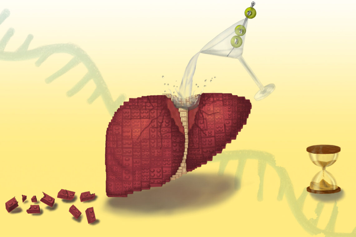
Long-term alcohol consumption causes a breakdown in the liver’s regenerative abilities, thanks to inflammation disrupting the protein-making process, Illinois researchers and collaborators found in a new study. Illustration by Deyasini Roy
Excessive alcohol consumption can disrupt the liver’s unique regenerative abilities by trapping cells in limbo between their functional and regenerative states, even after a patient stops drinking, researchers at University of Illinois Urbana-Champaign and collaborators at Duke University and the Chan Zuckerberg Biohub Chicago describe in a new study.
This in-between state is a result of inflammation disrupting how RNA is spliced during the protein-making process, the researchers found, providing scientists with new treatment pathways to explore for the deadly disease. The researchers published their findings in the journal Nature Communications.
The liver has a remarkable ability to regenerate itself after damage or partial removal. However, it loses that ability in patients with alcohol-associated liver disease — the leading cause of liver-related mortality worldwide, resulting in roughly 3 million deaths annually.
“We knew that the liver stops functioning and stops regenerating in patients with alcohol-related hepatitis and cirrhosis, even when a patient has discontinued consuming alcohol, but we didn’t know why,” said U. of I. biochemistry professor Auinash Kalsotra, who co-led the study with Duke University School of Medicine professor Anna Mae Diehl. “The only real life-saving treatment option once a patient reaches the liver failure stage in those diseases is transplantation. But if we understood why these livers were failing, maybe we could intervene.”
Both the Kalsotra and Diehl labs havestudied the molecular and cellular underpinnings of liver regeneration. Over the last five years, they found that in order to regenerate, liver cells reprogram their gene expression to revert to fetal-like progenitor cells, multiply and then reverse the process back to become mature functioning cells again. Armed with this knowledge, the group turned to the question of how those mechanisms were disrupted in alcohol-associated liver disease.
The researchers compared samples of healthy livers and samples of livers with alcohol-associated hepatitis or cirrhosis obtained from Johns Hopkins University Hospital through an initiative supported by the National Institute for Alcohol Abuse and Alcoholism, part of the National Institutes of Health.
Multiomic profiling of human alcohol-associated liver diseases

a Schematic summarizing single nuclear isolation from human liver samples and transcriptomics using multiomic and deep sequencing approaches. b rnaUMAP showing distinct cell populations identified from multiomic profiling at single-cell resolution from over 27,000 liver nuclei from 15 individuals (n = 5 each). c Distribution of various liver cells in unaffected and alcohol-associated liver disease (ALD) patients. AC and SAH showed an increased number of non-hepatocyte (NPC) cells. Non-directional Chi-squared test was performed cell numbers showed a p-value < 0.0001, indicating statistically significant change in cell populations depending on conditions. d Distribution of porto-central coordinate values in unaffected liver, AC, and SAH conditions. The histogram shows the frequency of cells at different values for each condition, followed by p-value from using two-sided Kolmogorov–Smirnov (K–S) test, which shows significant differences (p < 0.05) between unaffected and diseased distributions. e Cell type-specific changes in gene expression associated with ALD (examples shown in dot plot, left) and corresponding gene ontology analysis demonstrating affected processes (right). f Average Transcript per million (TPM) values of key pro-inflammatory cytokines as quantified from bulk RNA-seq. Correspoding Average TPM values are shown within each box of the heatmap. SAH severe alcohol-associated hepatitis, AC alcohol-associated cirrhosis.
The first thing the researchers noticed in diseased livers was that, although damaged cells had begun the process of reverting to the regenerative state, they did not complete the process and instead remained in transitional limbo.
“They are neither functional adult cells nor proliferative progenitor cells. Since they are not functioning, more pressure builds on the remaining cells. So they try to regenerate, and they’re all ending up in this unproductive quasi-progenitor state, and that’s what is causing liver failure,” said U. of I. graduate students Ullas Chembazhi and Sushant Bangru, the co-first authors of the study.
To figure out why the cells were getting stuck in this state, the team investigated which proteins were being made by the liver cells and, in turn, the RNA molecules carrying the instructions for those proteins from the DNA to the cell’s protein-building machinery.
While most studies focus only on the total amounts of RNA or protein in a cell, Kalsotra’s team used deep RNA sequencing technology and computational analyses to zoom in on the splicing of RNA fragments, a key step in stitching together different parts of genetic instructions to make proteins.
“In comparing the samples, we saw RNA was getting misspliced broadly in alcohol-related liver disease, across thousands of genes, and it was affecting major functions of proteins,” said Kalsotra, who also is affiliated with the Carl R. Woese Institute for Genomic Biology at Illinois.
The researchers found a possible driver of the RNA missplicing: Alcohol-damaged liver cells had a deficiency of the protein ESRP2, which binds to RNA to splice it properly.
“Proteins function at a very specific place in the cell, and that is directed by sequences within the protein that take the protein to that particular spot. We found that, in many cases, the sequence that dictates where the protein localizes within a cell was misspliced. That’s why it was important that we did the multiple analyses we did,” said Kalsotra, also a member of the Chan Zuckerberg Biohub Chicago. “There was the same amount of RNA and protein, but the protein was not at the right place to function. Due to missplicing, key proteins that are required for productive liver regeneration were getting stuck in the cytoplasm, when they needed to be in the nucleus.”
To verify that ESRP2 deficiency was a likely culprit, the researchers studied mice without the gene that produces ESRP2. They displayed similar liver damage and regeneration failure to that seen in patients with advanced alcohol-related hepatitis.
But why was ESRP2 missing from liver cells from patients with alcohol-related hepatitis? Upon investigation, the researchers found that liver support cells and immune cells, drawn to the liver tissue damaged by alcohol processing, released high amounts of inflammatory and growth factors. Those factors suppress ESRP2 production and activity.
To verify this finding, the researchers treated liver cell cultures with a molecule that inhibits the receptor for one of the inflammation-promoting factors. ESRP2 levels recovered and splicing activity was corrected, pointing to the pathway as a possible treatment target.
“I’m hopeful these findings will become a launching pad for future clinical studies. We can use these misspliced RNAs as diagnostic markers or develop treatments that can curb the inflammation. And if we can correct the splicing defects, then maybe we can improve recovery and restore damaged livers,” Kalsotra said.
Source – University of Illinois Urbana-Champaign


Long-term alcohol consumption causes a breakdown in the liver’s regenerative abilities, thanks to inflammation disrupting the protein-making process, Illinois researchers and collaborators found in a new study. Illustration by Deyasini Roy
Excessive alcohol consumption can disrupt the liver’s unique regenerative abilities by trapping cells in limbo between their functional and regenerative states, even after a patient stops drinking, researchers at University of Illinois Urbana-Champaign and collaborators at Duke University and the Chan Zuckerberg Biohub Chicago describe in a new study.
This in-between state is a result of inflammation disrupting how RNA is spliced during the protein-making process, the researchers found, providing scientists with new treatment pathways to explore for the deadly disease. The researchers published their findings in the journal Nature Communications.
The liver has a remarkable ability to regenerate itself after damage or partial removal. However, it loses that ability in patients with alcohol-associated liver disease — the leading cause of liver-related mortality worldwide, resulting in roughly 3 million deaths annually.
Both the Kalsotra and Diehl labs havestudied the molecular and cellular underpinnings of liver regeneration. Over the last five years, they found that in order to regenerate, liver cells reprogram their gene expression to revert to fetal-like progenitor cells, multiply and then reverse the process back to become mature functioning cells again. Armed with this knowledge, the group turned to the question of how those mechanisms were disrupted in alcohol-associated liver disease.
The researchers compared samples of healthy livers and samples of livers with alcohol-associated hepatitis or cirrhosis obtained from Johns Hopkins University Hospital through an initiative supported by the National Institute for Alcohol Abuse and Alcoholism, part of the National Institutes of Health.
Multiomic profiling of human alcohol-associated liver diseases
a Schematic summarizing single nuclear isolation from human liver samples and transcriptomics using multiomic and deep sequencing approaches. b rnaUMAP showing distinct cell populations identified from multiomic profiling at single-cell resolution from over 27,000 liver nuclei from 15 individuals (n = 5 each). c Distribution of various liver cells in unaffected and alcohol-associated liver disease (ALD) patients. AC and SAH showed an increased number of non-hepatocyte (NPC) cells. Non-directional Chi-squared test was performed cell numbers showed a p-value < 0.0001, indicating statistically significant change in cell populations depending on conditions. d Distribution of porto-central coordinate values in unaffected liver, AC, and SAH conditions. The histogram shows the frequency of cells at different values for each condition, followed by p-value from using two-sided Kolmogorov–Smirnov (K–S) test, which shows significant differences (p < 0.05) between unaffected and diseased distributions. e Cell type-specific changes in gene expression associated with ALD (examples shown in dot plot, left) and corresponding gene ontology analysis demonstrating affected processes (right). f Average Transcript per million (TPM) values of key pro-inflammatory cytokines as quantified from bulk RNA-seq. Correspoding Average TPM values are shown within each box of the heatmap. SAH severe alcohol-associated hepatitis, AC alcohol-associated cirrhosis.
The first thing the researchers noticed in diseased livers was that, although damaged cells had begun the process of reverting to the regenerative state, they did not complete the process and instead remained in transitional limbo.
To figure out why the cells were getting stuck in this state, the team investigated which proteins were being made by the liver cells and, in turn, the RNA molecules carrying the instructions for those proteins from the DNA to the cell’s protein-building machinery.
While most studies focus only on the total amounts of RNA or protein in a cell, Kalsotra’s team used deep RNA sequencing technology and computational analyses to zoom in on the splicing of RNA fragments, a key step in stitching together different parts of genetic instructions to make proteins.
The researchers found a possible driver of the RNA missplicing: Alcohol-damaged liver cells had a deficiency of the protein ESRP2, which binds to RNA to splice it properly.
To verify that ESRP2 deficiency was a likely culprit, the researchers studied mice without the gene that produces ESRP2. They displayed similar liver damage and regeneration failure to that seen in patients with advanced alcohol-related hepatitis.
But why was ESRP2 missing from liver cells from patients with alcohol-related hepatitis? Upon investigation, the researchers found that liver support cells and immune cells, drawn to the liver tissue damaged by alcohol processing, released high amounts of inflammatory and growth factors. Those factors suppress ESRP2 production and activity.
To verify this finding, the researchers treated liver cell cultures with a molecule that inhibits the receptor for one of the inflammation-promoting factors. ESRP2 levels recovered and splicing activity was corrected, pointing to the pathway as a possible treatment target.
Source – University of Illinois Urbana-Champaign
Chembazhi U, Bangru S, Dutta R, Das D, Peiffer B, Natua S, Toohill K, Leona A, Purwar I, Bhowmik A, Goyal Y, Sun Z, Diehl A, Kalsotra A. (2025) Dysregulated RNA splicing impairs regeneration in alcohol-associated liver disease. Nature Communications 16(1): 8049. [article]
Related Posts
Novel biosensor uses DNA ‘barcodes’ to uncover disease signals in cells
CellART extracts single-cell information from high-resolution spatial transcriptomics
Cell2space restores spatial context to single-cell RNA sequencing data
Urine test may improve bladder cancer diagnosis and guide treatment decisions
DeCoRE – a computational method to resolve RNA structural heterogeneity from RNA structure probing data by direct RNA sequencing
Tracing 700 million years of cell evolution
How should biological replicates be handled in long-read RNA sequencing?
RNA-Seq reveals a new way the developing heart controls its growth
The Human RNome Project Consortium seeks to publish first-ever complete sequence of the body’s RNA
scFlowReport brings multiple single-cell RNA sequencing analyses into one workflow
Long-read RNA sequencing reveals how neurons choose alternative RNA endings
DeOPUS improves cell-type estimates from bulk RNA sequencing
ResolVI improves accuracy in spatial transcriptomics analysis
RNA sequencing reveals transcriptional allele differences in prostate cancer stem cells
Unmasking “zombie cells” in aging tissue with an AI-powered barcode
RNA variant imaging for cancer diagnosis
Autism-risk mutations reveal two opposing patterns of brain gene activity
A real-data approach to evaluating RNA transcript analysis methods
Detection and sequencing of Ap2N-capped RNAs in human cells
Single-cell RNA sequencing provides a closer look at the Aedes aegypti midgut
Stay Connected
Submit a Post to the Blog
Recent Posts
Subscribe to the RNA-Seq Blog
RNA-Seq Products & Services