

Small changes in DNA can have a big impact on how cells function. In cancer, mutations often alter how RNA molecules fold and behave. These structural changes can affect how RNA interacts with drugs, especially small molecules designed to bind RNA targets. Until now, it has been difficult to systematically measure how individual mutations influence these interactions.
Researchers at Kyoto University in Japan have developed a new method to address this challenge. Their technique, called BIVID-MaP, allows scientists to detect how small molecules bind to RNA and how this binding changes when mutations are present.
Modification-induced RT deletions to detect RNA-small molecule interactions
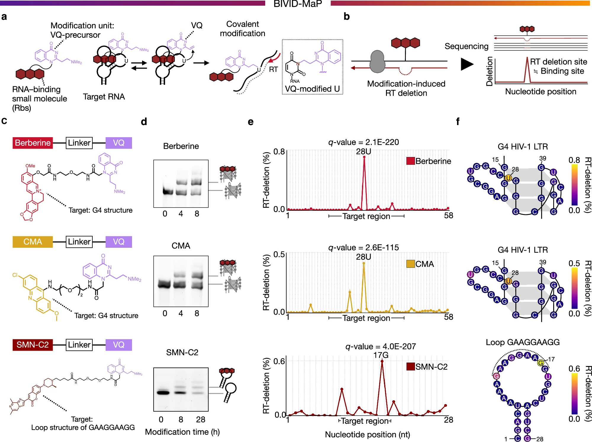
a, b Overview of BIVID-MaP. a Overview of binding-dependent modification by Rbs-VQ. Rbs (binding unit) binds to target RNA structures and then VQ (modification unit) covalently modifies the base near target RNA structures. b The binding site is detected by reverse transcription deletion (RT deletion) caused by binding-dependent modifications. c Chemical structures of alkylating modifiers used to probe RNA binding by berberine, CMA, and SMN-C2. d Gel shift assays showing the covalent modification by each modifier on the target RNA sequence. Berberine and CMA target G4 HIV-1 LTR and SMN-C2 targets Loop GAAGGAAGG. For visualization, SYBR Gold staining was used for the G4 HIV-1 LTR, and 5′-FAM labeling was used for the Loop GAAGGAAGG. From bottom to top, the gel shift bands show unmodified RNA, a 1:1 covalent complex (one modifier per RNA) and a 1:2 covalent complex (two modifiers per RNA) (See Supplementary Note). Representative data are shown from technical triplicate. e Modification-derived RT deletion frequency (RT-deletion), calculated as the deletion percentage with modifier minus the deletion percentage without modifier. Plots show RT-deletion at each nucleotide of the target binding site for each small molecule. The x-axis indicates nucleotide positions in G4 HIV-1 LTR for berberine and CMA, and in Loop GAAGGAAGG for SMN-C2. The target region is highlighted below each graph. f Model structures of the G-quadruplex in the HIV-1 LTR G4 and of the hairpin loop in Loop GAAGGAAGG, annotated with the RT-deletion (%) observed at each nucleotide. Target region of Loop GAAGGAAGG is highlighted with a dotted line. BIVID-MaP was performed in two independent experiments (n = 2).
The method works by combining chemical modification of RNA with sequencing based detection. When a small molecule binds to RNA, it triggers changes that can be captured during reverse transcription. These changes appear as deletions in the sequencing data, which can then be mapped back to specific regions of the RNA. Using RNA sequencing, the researchers can identify where and how strongly small molecules interact with RNA molecules across many different variants.
A key focus of the study was on G-quadruplex structures, which are unique folded RNA configurations known to play roles in gene regulation. The team examined how single nucleotide changes, such as those found in cancer, influence the ability of a small molecule to bind these structures.
The results showed that even a single mutation can significantly increase or decrease how strongly a drug binds to RNA. Some cancer associated mutations altered the structure of RNA in ways that either enhanced or disrupted small molecule interactions.
This finding is important because it suggests that RNA targeted drugs may behave differently depending on a patient’s specific genetic mutations. Understanding these differences could help researchers design more precise therapies that account for individual variation.
The BIVID-MaP approach provides a new way to study RNA structure and drug interactions at high resolution. By linking RNA sequencing data with chemical binding information, the method opens the door to more personalized approaches in drug development, especially for diseases like cancer where mutations play a central role.
Availability – Custom scripts for classifying sequencing reads and calculating RT deletions per variant are available on GitHub with instructions for their use (https://github.com/BIVID-MaP/BIVID_MaP2025)
Miyashita E, Onizuka K, Chen Y, Yoshida H, Hatayama H, Ishikawa S, Yan P, Hasegawa T, Ozawa M, Maeta K, Nagatsugi F, Saito H, Komatsu K R. (2026) Systematic identification of variant-specific RNA structure-small molecule interactions exemplified by RNA G-quadruplexes. Nature Communications 17(1): 2243. [article]
Related Posts






Small changes in DNA can have a big impact on how cells function. In cancer, mutations often alter how RNA molecules fold and behave. These structural changes can affect how RNA interacts with drugs, especially small molecules designed to bind RNA targets. Until now, it has been difficult to systematically measure how individual mutations influence these interactions.
Researchers at Kyoto University in Japan have developed a new method to address this challenge. Their technique, called BIVID-MaP, allows scientists to detect how small molecules bind to RNA and how this binding changes when mutations are present.
Modification-induced RT deletions to detect RNA-small molecule interactions
a, b Overview of BIVID-MaP. a Overview of binding-dependent modification by Rbs-VQ. Rbs (binding unit) binds to target RNA structures and then VQ (modification unit) covalently modifies the base near target RNA structures. b The binding site is detected by reverse transcription deletion (RT deletion) caused by binding-dependent modifications. c Chemical structures of alkylating modifiers used to probe RNA binding by berberine, CMA, and SMN-C2. d Gel shift assays showing the covalent modification by each modifier on the target RNA sequence. Berberine and CMA target G4 HIV-1 LTR and SMN-C2 targets Loop GAAGGAAGG. For visualization, SYBR Gold staining was used for the G4 HIV-1 LTR, and 5′-FAM labeling was used for the Loop GAAGGAAGG. From bottom to top, the gel shift bands show unmodified RNA, a 1:1 covalent complex (one modifier per RNA) and a 1:2 covalent complex (two modifiers per RNA) (See Supplementary Note). Representative data are shown from technical triplicate. e Modification-derived RT deletion frequency (RT-deletion), calculated as the deletion percentage with modifier minus the deletion percentage without modifier. Plots show RT-deletion at each nucleotide of the target binding site for each small molecule. The x-axis indicates nucleotide positions in G4 HIV-1 LTR for berberine and CMA, and in Loop GAAGGAAGG for SMN-C2. The target region is highlighted below each graph. f Model structures of the G-quadruplex in the HIV-1 LTR G4 and of the hairpin loop in Loop GAAGGAAGG, annotated with the RT-deletion (%) observed at each nucleotide. Target region of Loop GAAGGAAGG is highlighted with a dotted line. BIVID-MaP was performed in two independent experiments (n = 2).
The method works by combining chemical modification of RNA with sequencing based detection. When a small molecule binds to RNA, it triggers changes that can be captured during reverse transcription. These changes appear as deletions in the sequencing data, which can then be mapped back to specific regions of the RNA. Using RNA sequencing, the researchers can identify where and how strongly small molecules interact with RNA molecules across many different variants.
A key focus of the study was on G-quadruplex structures, which are unique folded RNA configurations known to play roles in gene regulation. The team examined how single nucleotide changes, such as those found in cancer, influence the ability of a small molecule to bind these structures.
The results showed that even a single mutation can significantly increase or decrease how strongly a drug binds to RNA. Some cancer associated mutations altered the structure of RNA in ways that either enhanced or disrupted small molecule interactions.
This finding is important because it suggests that RNA targeted drugs may behave differently depending on a patient’s specific genetic mutations. Understanding these differences could help researchers design more precise therapies that account for individual variation.
The BIVID-MaP approach provides a new way to study RNA structure and drug interactions at high resolution. By linking RNA sequencing data with chemical binding information, the method opens the door to more personalized approaches in drug development, especially for diseases like cancer where mutations play a central role.
Availability – Custom scripts for classifying sequencing reads and calculating RT deletions per variant are available on GitHub with instructions for their use (https://github.com/BIVID-MaP/BIVID_MaP2025)
Miyashita E, Onizuka K, Chen Y, Yoshida H, Hatayama H, Ishikawa S, Yan P, Hasegawa T, Ozawa M, Maeta K, Nagatsugi F, Saito H, Komatsu K R. (2026) Systematic identification of variant-specific RNA structure-small molecule interactions exemplified by RNA G-quadruplexes. Nature Communications 17(1): 2243. [article]
Related Posts
Recent Posts
 Scientists identify the neuroblastoma cells that signal poor outcomes
 GARAGE improves rare cell detection in single-cell RNA sequencing analysis using AI
 Machine learning reveals how RNA splicing is regulated in individual cell types
 Illumina releases SpliceAI2 to help advance rare disease research
 Postdoctoral Fellow – Long-Read RNA Methods Development for Neurodegeneration
 Novel biosensor uses DNA ‘barcodes’ to uncover disease signals in cells
RNA-Seq Products & Services



Stay Connected