Scientists at the Icahn School of Medicine at Mount Sinai, in partnership with the Multiple Myeloma Research Foundation (MMRF) and in collaboration with leading institutions across the country, have helped generate the largest single-cell immune cell atlas of the bone marrow in patients with multiple myeloma, a blood cancer that, while treatable, remains incurable. The findings, published in Nature Cancer, provide unprecedented insight on immune dysfunction in myeloma and could lead to improved tools for predicting which patients are at higher risk of relapse after treatment.

The study analyzed nearly 1.5 million immune cells from the bone marrow of 335 newly diagnosed patients in the MMRF’s CoMMpass study dataset, using a cutting-edge technique called single-cell RNA sequencing. This approach allowed researchers to examine the molecular features of individual cells, revealing previously hidden patterns in how the immune system behaves in the presence of multiple myeloma.
“Currently, doctors rely heavily on the genetic features of cancer cells to estimate how aggressive their disease is,” said co-senior author Sacha Gnjatic, PhD, Professor of Immunology and Immunotherapy at the Mount Sinai Tisch Cancer Center. “Our research shows that the immune cells surrounding the tumor are just as important in determining how the disease progresses, and this could lead to earlier, more targeted treatment strategies.”
“The scale and maturity of the MMRF’s CoMMpass study and the breadth of this multi-center collaboration highlights the importance of creating ‘big science’ teams,” added George Mulligan, PhD, co-senior author and Chief Scientific Officer at the MMRF.
Overview of the Immune Atlas design, workflow and participant characteristics
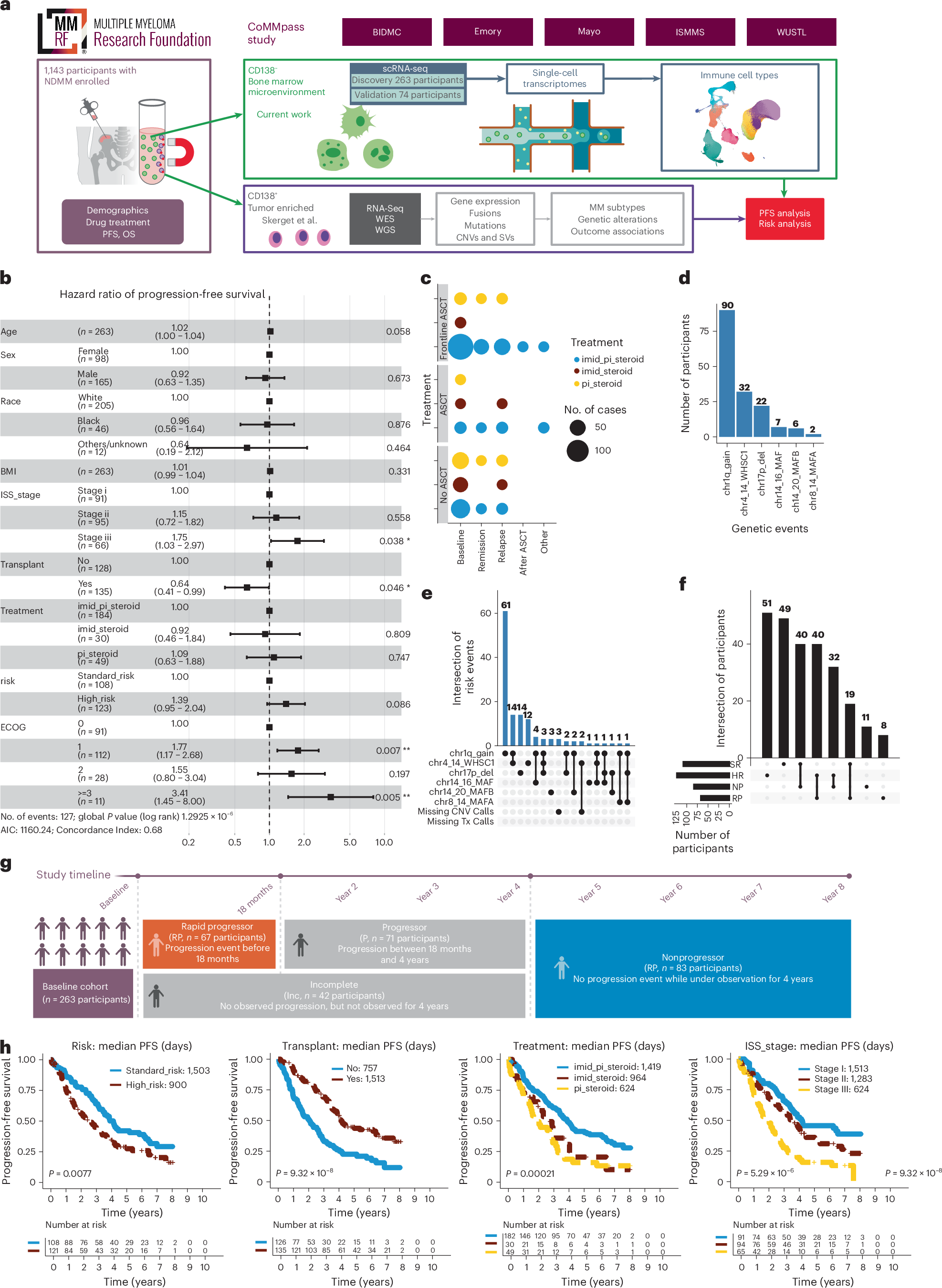
a, Overview of the Immune Atlas study design, discovery (nparticipants = 263) and validation (nparticipants = 74) participant cohorts, sample processing and analysis workflow. b, Clinical characteristics of participants (nparticipants = 263) in the discovery cohort. The forest plot illustrates the effect of various clinical features on PFS. Error bars display the 95% confidence interval (CI). c, Dot plot depicting the cross-section of samples based on ASCT and frontline treatment, where the dot size indicates the number of participants and dot color indicates the type of treatment regimen. d, Bar chart showing the total number of participants with each of the six genetic events used for risk classification. e, UpSet plot showing the distribution of the major cytogenetic abnormalities comprising the Davies-based HR myeloma definition. f, UpSet plot showing the intersection of participants categorized as SR or HR and NP or RP at baseline. g, Overview of progression group categorization and study design for the discovery cohort. The participants with a progression event within the first 18 months following therapy were classified as RPs (nparticipants = 67). Participants with durable remission or no observed progression for at least 4 years were classified as NPs (nparticipants = 83). Participants with a progression event between 18 months and 4 years were classified as RPs (nparticipants = 71). The participants who exited the study before 4 years of disease diagnosis without experiencing a progression event were classified as Inc (n = 42). h, Kaplan–Meier curves display the survival analysis for participants categorized on the basis of risk stratification (HR versus SR), transplant as a frontline treatment, treatment type and ISS staging. Participants lacking ISS stage information at baseline or WGS information for cytogenetic risk stratification were omitted from the relevant figure panels. The P values were estimated using a log-rank test.
The team discovered that some patients who relapsed quickly after initial therapy had distinct immune cell populations in their bone marrow at the time of diagnosis. Notably, a subset of T cells (immune cells typically responsible for attacking tumors) appeared to show a state of immunosenescence, meaning they were present but not functioning effectively and contributing to immune suppression. In addition, the study uncovered patterns of communication between immune cells and cancer cells that may support tumor growth.
These insights suggest that the immune system’s response to myeloma could be used as a predictive tool, offering a complementary layer to traditional genetic testing. While the current technology used in the study is primarily for research, the investigators hope the findings will inform the development of simple, broadly available tests in the future.
“This work not only provides new biological insights, but also lays the groundwork for future discoveries,” Dr. Gnjatic said. “It could help researchers around the world better understand how the immune system interacts with cancer and ultimately, help improve outcomes for patients.”
The MMRF designed and funded this collaborative research, which involved equal contribution from scientists at Mount Sinai, Emory University, Beth Israel Deaconess Medical Center, Washington University in St. Louis’s Siteman Cancer Center, the Mayo Clinic, and the MMRF. Mount Sinai’s Edgar Gonzalez-Kozlova, PhD, was co-first author along with other lead data analysts from the six institutions, while other important contributors from Mount Sinai included Seunghee Kim-Schulze, PhD, the lead for data generation, and Hearn J. Cho, MD/PhD, a Mount Sinai myeloma oncologist and researcher who is also Chief Medical Officer at the MMRF.
The large patient sample size and robust clinical annotations, enabled by the MMRF’s extensive biorepository and 12 years of patient data, make this one of the most comprehensive resources of its kind. This report will significantly contribute to our understanding of immune function in multiple myeloma.
Source – Icahn School of Medicine at Mount Sinai


Scientists at the Icahn School of Medicine at Mount Sinai, in partnership with the Multiple Myeloma Research Foundation (MMRF) and in collaboration with leading institutions across the country, have helped generate the largest single-cell immune cell atlas of the bone marrow in patients with multiple myeloma, a blood cancer that, while treatable, remains incurable. The findings, published in Nature Cancer, provide unprecedented insight on immune dysfunction in myeloma and could lead to improved tools for predicting which patients are at higher risk of relapse after treatment.
The study analyzed nearly 1.5 million immune cells from the bone marrow of 335 newly diagnosed patients in the MMRF’s CoMMpass study dataset, using a cutting-edge technique called single-cell RNA sequencing. This approach allowed researchers to examine the molecular features of individual cells, revealing previously hidden patterns in how the immune system behaves in the presence of multiple myeloma.
Overview of the Immune Atlas design, workflow and participant characteristics
a, Overview of the Immune Atlas study design, discovery (nparticipants = 263) and validation (nparticipants = 74) participant cohorts, sample processing and analysis workflow. b, Clinical characteristics of participants (nparticipants = 263) in the discovery cohort. The forest plot illustrates the effect of various clinical features on PFS. Error bars display the 95% confidence interval (CI). c, Dot plot depicting the cross-section of samples based on ASCT and frontline treatment, where the dot size indicates the number of participants and dot color indicates the type of treatment regimen. d, Bar chart showing the total number of participants with each of the six genetic events used for risk classification. e, UpSet plot showing the distribution of the major cytogenetic abnormalities comprising the Davies-based HR myeloma definition. f, UpSet plot showing the intersection of participants categorized as SR or HR and NP or RP at baseline. g, Overview of progression group categorization and study design for the discovery cohort. The participants with a progression event within the first 18 months following therapy were classified as RPs (nparticipants = 67). Participants with durable remission or no observed progression for at least 4 years were classified as NPs (nparticipants = 83). Participants with a progression event between 18 months and 4 years were classified as RPs (nparticipants = 71). The participants who exited the study before 4 years of disease diagnosis without experiencing a progression event were classified as Inc (n = 42). h, Kaplan–Meier curves display the survival analysis for participants categorized on the basis of risk stratification (HR versus SR), transplant as a frontline treatment, treatment type and ISS staging. Participants lacking ISS stage information at baseline or WGS information for cytogenetic risk stratification were omitted from the relevant figure panels. The P values were estimated using a log-rank test.
The team discovered that some patients who relapsed quickly after initial therapy had distinct immune cell populations in their bone marrow at the time of diagnosis. Notably, a subset of T cells (immune cells typically responsible for attacking tumors) appeared to show a state of immunosenescence, meaning they were present but not functioning effectively and contributing to immune suppression. In addition, the study uncovered patterns of communication between immune cells and cancer cells that may support tumor growth.
These insights suggest that the immune system’s response to myeloma could be used as a predictive tool, offering a complementary layer to traditional genetic testing. While the current technology used in the study is primarily for research, the investigators hope the findings will inform the development of simple, broadly available tests in the future.
The MMRF designed and funded this collaborative research, which involved equal contribution from scientists at Mount Sinai, Emory University, Beth Israel Deaconess Medical Center, Washington University in St. Louis’s Siteman Cancer Center, the Mayo Clinic, and the MMRF. Mount Sinai’s Edgar Gonzalez-Kozlova, PhD, was co-first author along with other lead data analysts from the six institutions, while other important contributors from Mount Sinai included Seunghee Kim-Schulze, PhD, the lead for data generation, and Hearn J. Cho, MD/PhD, a Mount Sinai myeloma oncologist and researcher who is also Chief Medical Officer at the MMRF.
The large patient sample size and robust clinical annotations, enabled by the MMRF’s extensive biorepository and 12 years of patient data, make this one of the most comprehensive resources of its kind. This report will significantly contribute to our understanding of immune function in multiple myeloma.
Source – Icahn School of Medicine at Mount Sinai
Pilcher W C, Yao L, Gonzalez-Kozlova E, Pita-Juarez Y, Karagkouni D, Acharya C R, Michaud M E, Hamilton M, Nanda S, Song Y, Sato K, Wang J T, Satpathy S, Ma Y, Schulman J, D’Souza D, Jayasinghe R G, Ohlstrom D, Ferguson K E, Cheloni G, Bakhtiari M, Pabustan N, Nie K, Foltz J A, Saldarriaga I, Alaaeldin R, Lepisto E, Chen R, Fiala M A, Thomas B E, Cook A, Santos J V D, I-Ling C, Figueiredo I, Fortier J, Slade M, Oh S T, Rettig M P, Anderson E, Li Y, Dasari S, Strausbauch M A, Simon V A, Radkevich E, Rahman A H, Chen Z, Lagana A, DiPersio J F, Rosenblatt J, Kim-Schulze S, Lonial S, Kumar S, Bhasin S S, Kourelis T, Dhodapkar M V, Vij R, Avigan D, Cho H J, Mulligan G, Ding L, Gnjatic S, Vlachos I S, Bhasin M. (2026) A single-cell atlas characterizes dysregulation of the bone marrow immune microenvironment associated with outcomes in multiple myeloma. Nature Cancer [Epub ahead of print]. [article]
Related Posts
Scientists identify the neuroblastoma cells that signal poor outcomes
GARAGE improves rare cell detection in single-cell RNA sequencing analysis using AI
Novel biosensor uses DNA ‘barcodes’ to uncover disease signals in cells
CellART extracts single-cell information from high-resolution spatial transcriptomics
Cell2space restores spatial context to single-cell RNA sequencing data
Urine test may improve bladder cancer diagnosis and guide treatment decisions
DeCoRE – a computational method to resolve RNA structural heterogeneity from RNA structure probing data by direct RNA sequencing
Tracing 700 million years of cell evolution
How should biological replicates be handled in long-read RNA sequencing?
RNA-Seq reveals a new way the developing heart controls its growth
The Human RNome Project Consortium seeks to publish first-ever complete sequence of the body’s RNA
scFlowReport brings multiple single-cell RNA sequencing analyses into one workflow
Long-read RNA sequencing reveals how neurons choose alternative RNA endings
DeOPUS improves cell-type estimates from bulk RNA sequencing
ResolVI improves accuracy in spatial transcriptomics analysis
RNA sequencing reveals transcriptional allele differences in prostate cancer stem cells
Unmasking “zombie cells” in aging tissue with an AI-powered barcode
RNA variant imaging for cancer diagnosis
Autism-risk mutations reveal two opposing patterns of brain gene activity
A real-data approach to evaluating RNA transcript analysis methods
Stay Connected
Submit a Post to the Blog
Recent Posts
Subscribe to the RNA-Seq Blog
RNA-Seq Products & Services