Researchers at Tel Aviv University’s Gray Faculty of Medical & Health Sciences conducted a series of experiments using advanced technologies, focusing on the tumor microenvironment in advanced stages of breast cancer. They found that immune system cells called neutrophils are recruited by the tumor environment and support tumor progression in the later stages of the disease.
The researchers stated: “In light of our findings, we believe that neutrophils, which are unique to the tumor environment, may serve as targets for the development of new drugs and as biomarkers for advanced-stage breast cancer.”
Dr. Cohen explained: “In our lab, we study cell-to-cell communication at the single-cell level using an innovative single-cell RNA sequencing method, combined with bioinformatics analyses. Additionally, we employ a unique method for RNA sequencing of pairs of interacting cells, allowing us to study the communication itself. In the current study, we focused on the intercellular communication between breast cancer cells and neutrophils in the tumor microenvironment.”
Using a mouse model, the researchers collected breast tissue samples at four different stages: at 10 days old, 3 weeks old—corresponding to breast development in adolescent girls—as well as from healthy adult mice and adult mice with breast cancer. Single-cell RNA sequencing was performed on all samples, and the findings were analyzed using advanced bioinformatics methods. Through this approach, two types of cells were identified as predominantly characterizing advanced-stage cancer tissues: neutrophil immune cells adjacent to cancer cells, and endothelial cells, which among other roles, promote blood vessel development.
Thorough characterization of mammary gland immune and nonimmune lineages
during physiological development and carcinogenesis
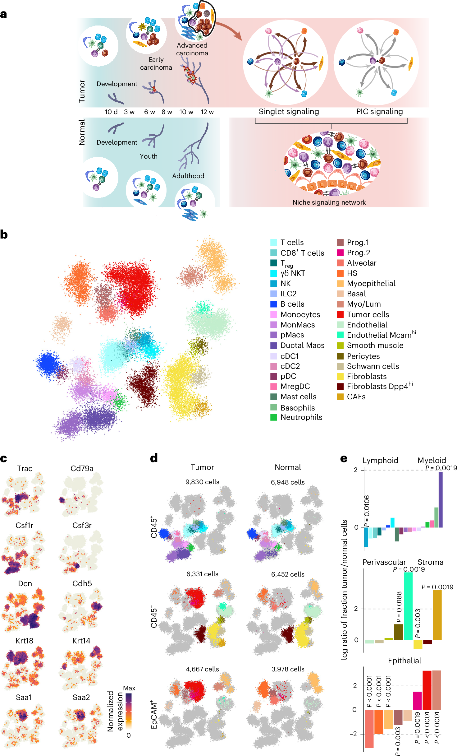
a, Experimental design. Mammary gland samples were collected at different time points from PyMT+ (tumor) and PyMT− (normal) mice. scRNA-seq and PIC-seq analysis enabled investigation of the molecular signaling in the tumor niche. 10 d: mammary glands from 5–6 neonates were pooled in each biological replicate, PyMT− n = 3; PyMT+ n = 3; 3 w: mammary glands from three mice were pooled, PyMT− n = 4; PyMT+ n = 5; 6–8 w: mammary glands from two mice were pooled, PyMT− n = 5; PyMT+ n = 5; 10–12 w: mammary glands from 1–2 mice were pooled, PyMT− n = 5; PyMT+ n = 11; Supplementary Table 1. b, A two-dimensional projection of 38,206 single cells, grouped into 683 metacells, based on the neighbor graph created by the MetaCell package. Cells are colored by annotation based on hallmark gene expression. c, A two-dimensional projection of key cell-type-specific marker gene log-normalized expression in all cells. d, Two-dimensional projection maps highlighting cells originating from tumor and normal mice and from three different sorted populations, CD45+, CD45− and EpCAM+. e, log ratio between the population fractions of each cell type in tumor/normal mammary glands, demonstrating positive or negative enrichment in tumor versus normal samples. The fraction was calculated as the number of cells from the relevant FACS gate that belong to each annotation, averaged first across biological replicates in each time point then across time points. P values were determined using a two-way analysis of variance (ANOVA) followed by Tukey’s post hoc test for multiple comparisons. The test was run before averaging across time points, comparing the population fraction across time points and conditions.
Dr. Camargo and Ori Moskowitz added: “From the findings, we inferred that neutrophils play a role in breast cancer. To explore this further, we examined their molecular communication—that is, the signals they send and receive within their environment. In doing so, we uncovered a complex process that supports tumor growth: cancer cells stimulate macrophages (immune system cells located in the milk ducts of the breast) to recruit neutrophils into the tumor microenvironment. Once there, the neutrophils physically interact with the cancer cells, leading to the secretion of substances that enhance the cancer’s ability to spread and invade additional tissues and increase the formation of blood vessels that nourish the tumor.”
The findings were validated by two additional tests: First, the researchers showed that silencing neutrophils in mice with advanced breast cancer significantly reduced the proliferation of blood vessels and tumor cells. Second, they found that the survival rate of women with late-stage breast cancer (Stages 3 and 4) decreased the more they expressed molecular signatures of neutrophils and their interactions with cancer cells.
Dr. Cohen concluded: “In this study, we uncovered an important mechanism in the development of breast cancer. We found that neutrophils—immune system cells recruited into the tumor environment—play a central role in supporting cancer progression. These findings have dual significance for breast cancer diagnosis and treatment: neutrophils and the signals they emit may serve both as targets for the development of new drugs and as biomarkers for the advanced stages of the disease.”
Source– Eurekalert


Researchers at Tel Aviv University’s Gray Faculty of Medical & Health Sciences conducted a series of experiments using advanced technologies, focusing on the tumor microenvironment in advanced stages of breast cancer. They found that immune system cells called neutrophils are recruited by the tumor environment and support tumor progression in the later stages of the disease.
Using a mouse model, the researchers collected breast tissue samples at four different stages: at 10 days old, 3 weeks old—corresponding to breast development in adolescent girls—as well as from healthy adult mice and adult mice with breast cancer. Single-cell RNA sequencing was performed on all samples, and the findings were analyzed using advanced bioinformatics methods. Through this approach, two types of cells were identified as predominantly characterizing advanced-stage cancer tissues: neutrophil immune cells adjacent to cancer cells, and endothelial cells, which among other roles, promote blood vessel development.
Thorough characterization of mammary gland immune and nonimmune lineages
during physiological development and carcinogenesis
a, Experimental design. Mammary gland samples were collected at different time points from PyMT+ (tumor) and PyMT− (normal) mice. scRNA-seq and PIC-seq analysis enabled investigation of the molecular signaling in the tumor niche. 10 d: mammary glands from 5–6 neonates were pooled in each biological replicate, PyMT− n = 3; PyMT+ n = 3; 3 w: mammary glands from three mice were pooled, PyMT− n = 4; PyMT+ n = 5; 6–8 w: mammary glands from two mice were pooled, PyMT− n = 5; PyMT+ n = 5; 10–12 w: mammary glands from 1–2 mice were pooled, PyMT− n = 5; PyMT+ n = 11; Supplementary Table 1. b, A two-dimensional projection of 38,206 single cells, grouped into 683 metacells, based on the neighbor graph created by the MetaCell package. Cells are colored by annotation based on hallmark gene expression. c, A two-dimensional projection of key cell-type-specific marker gene log-normalized expression in all cells. d, Two-dimensional projection maps highlighting cells originating from tumor and normal mice and from three different sorted populations, CD45+, CD45− and EpCAM+. e, log ratio between the population fractions of each cell type in tumor/normal mammary glands, demonstrating positive or negative enrichment in tumor versus normal samples. The fraction was calculated as the number of cells from the relevant FACS gate that belong to each annotation, averaged first across biological replicates in each time point then across time points. P values were determined using a two-way analysis of variance (ANOVA) followed by Tukey’s post hoc test for multiple comparisons. The test was run before averaging across time points, comparing the population fraction across time points and conditions.
The findings were validated by two additional tests: First, the researchers showed that silencing neutrophils in mice with advanced breast cancer significantly reduced the proliferation of blood vessels and tumor cells. Second, they found that the survival rate of women with late-stage breast cancer (Stages 3 and 4) decreased the more they expressed molecular signatures of neutrophils and their interactions with cancer cells.
Source– Eurekalert
Camargo S, Moskowitz O, Giladi A, Levinson M, Balaban R, Gola S, Raizman A, Lipczyc K, Richter A, Keren-Khadmy N, Barboy O, Dugach Y, Carmi Y, Sonnenblick A, Cohen M. (2025) Neutrophils physically interact with tumor cells to form a signaling niche promoting breast cancer aggressiveness. Nat Cancer 6(3):540–558. [article]
Related Posts
Novel biosensor uses DNA ‘barcodes’ to uncover disease signals in cells
CellART extracts single-cell information from high-resolution spatial transcriptomics
Cell2space restores spatial context to single-cell RNA sequencing data
Urine test may improve bladder cancer diagnosis and guide treatment decisions
DeCoRE – a computational method to resolve RNA structural heterogeneity from RNA structure probing data by direct RNA sequencing
Tracing 700 million years of cell evolution
How should biological replicates be handled in long-read RNA sequencing?
RNA-Seq reveals a new way the developing heart controls its growth
The Human RNome Project Consortium seeks to publish first-ever complete sequence of the body’s RNA
scFlowReport brings multiple single-cell RNA sequencing analyses into one workflow
Long-read RNA sequencing reveals how neurons choose alternative RNA endings
DeOPUS improves cell-type estimates from bulk RNA sequencing
ResolVI improves accuracy in spatial transcriptomics analysis
RNA sequencing reveals transcriptional allele differences in prostate cancer stem cells
Unmasking “zombie cells” in aging tissue with an AI-powered barcode
RNA variant imaging for cancer diagnosis
Autism-risk mutations reveal two opposing patterns of brain gene activity
A real-data approach to evaluating RNA transcript analysis methods
Detection and sequencing of Ap2N-capped RNAs in human cells
Single-cell RNA sequencing provides a closer look at the Aedes aegypti midgut
Stay Connected
Submit a Post to the Blog
Recent Posts
Subscribe to the RNA-Seq Blog
RNA-Seq Products & Services