

Study presents analytical workflow linking gene expression with measurable cell shape and size, enabling validation of transcriptional states against imaging data
A research team at St. Jude Children’s Research Hospital, has introduced a downstream analytical framework for STAMP (Single-cell Transcriptomics Analysis and Multimodal Profiling), an emerging technology that preserves cell morphology alongside gene and protein expression. In a mini review published in EXO – Beyond the Cell, the team demonstrates how STAMP data can be analyzed using standard Python-based single-cell tools to link molecular profiles directly with image-derived phenotypes—such as cell area, shape, and marker localization.
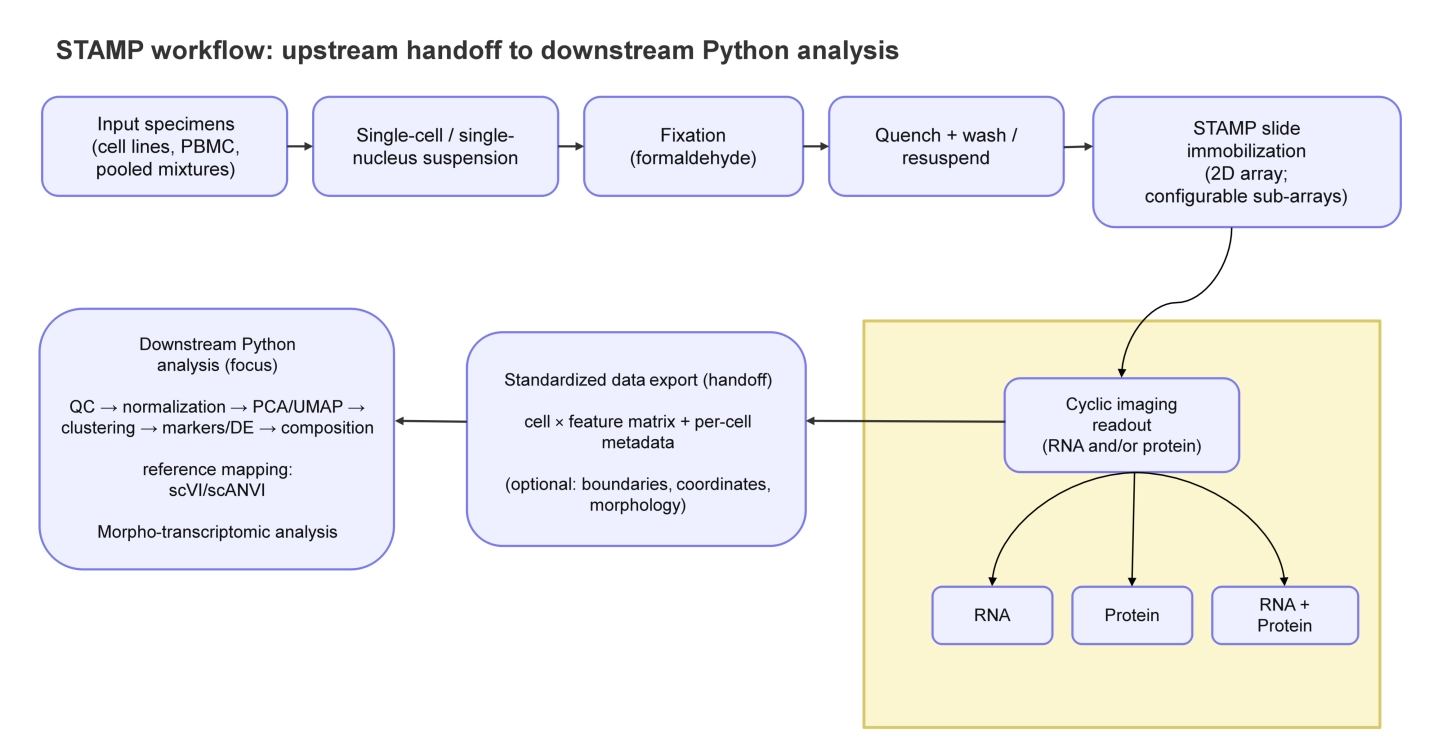
Dissociated cells or nuclei are immobilized on a slide, imaged (RNA +/- protein), and exported as an analysis-ready cell × feature matrix linked to per-cell morphology and coordinates. Downstream Python analysis applies standard single-cell steps (QC, normalization, embedding/latent modeling, clustering/label transfer, morpho-transcriptomic analysis, and interpretation), with results traceable back to the images. STAMP: single-cell transcriptomics analysis and multimodal profiling; QC: quality control; PBMC: peripheral blood mononuclear cell; PCA: principal component analysis; UMAP: Uniform Manifold Approximation and Projection; DE: differential expression; scVI: single-cell variational inference; scANVI: single-cell ANnotation using variational inference.
Unlike conventional single-cell RNA sequencing, which requires tissue dissociation and loses spatial context, STAMP immobilizes dissociated cells or nuclei on a slide, enabling high-content imaging before molecular readout. This preserves per-cell visual characteristics while allowing high-plex transcript and protein measurement. The result is a unified dataset where each cell is represented by both its gene expression vector and its morphological features.
The team validated a complete computational pipeline using a publicly available pooled mixture of three cancer cell lines (LNCaP, MCF7, and SKBR3) as a test case. They applied quality control informed by both molecular and imaging data, normalized expression, performed dimensionality reduction, and used semi-supervised label transfer (scVI and scANVI) to project unlabeled mixture cells onto pure reference cell line embeddings. The method recovered expected cell-type identities with high confidence and revealed that ambiguous or low-confidence assignments were extremely rare (0.095% of cells had posterior probability <0.8).
A key innovation highlighted in the work is morpho-transcriptomic coupling—explicitly testing whether transcriptionally inferred states correlate with co-measured morphology. For example, the team showed that a G2/M proliferation module score correlated with cell area in a cell-line–dependent manner, with effect sizes ranging from r = −0.18 to r = 0.19. This approach transforms morphology from a qualitative illustration into a measurable covariate that can confirm or challenge transcript-based conclusions.
The team emphasize that STAMP does not preserve native tissue geography (unlike intact-section spatial transcriptomics), but it offers scalability, experimental flexibility, and the unique ability to ground molecular inferences in directly imaged cellular phenotypes. The workflow is compatible with mainstream Python single-cell ecosystems such as Scanpy, scVI, and scANVI.
Source – Eurekalert
Poudel S, Dezem FS, Martelotto LG, Plummer JT, Green DR. (2026) Democratizing the spatial view: STAMP technology from an analytical perspective. EXO 1:202608. [article]
Related Posts






Study presents analytical workflow linking gene expression with measurable cell shape and size, enabling validation of transcriptional states against imaging data
A research team at St. Jude Children’s Research Hospital, has introduced a downstream analytical framework for STAMP (Single-cell Transcriptomics Analysis and Multimodal Profiling), an emerging technology that preserves cell morphology alongside gene and protein expression. In a mini review published in EXO – Beyond the Cell, the team demonstrates how STAMP data can be analyzed using standard Python-based single-cell tools to link molecular profiles directly with image-derived phenotypes—such as cell area, shape, and marker localization.
Dissociated cells or nuclei are immobilized on a slide, imaged (RNA +/- protein), and exported as an analysis-ready cell × feature matrix linked to per-cell morphology and coordinates. Downstream Python analysis applies standard single-cell steps (QC, normalization, embedding/latent modeling, clustering/label transfer, morpho-transcriptomic analysis, and interpretation), with results traceable back to the images. STAMP: single-cell transcriptomics analysis and multimodal profiling; QC: quality control; PBMC: peripheral blood mononuclear cell; PCA: principal component analysis; UMAP: Uniform Manifold Approximation and Projection; DE: differential expression; scVI: single-cell variational inference; scANVI: single-cell ANnotation using variational inference.
Unlike conventional single-cell RNA sequencing, which requires tissue dissociation and loses spatial context, STAMP immobilizes dissociated cells or nuclei on a slide, enabling high-content imaging before molecular readout. This preserves per-cell visual characteristics while allowing high-plex transcript and protein measurement. The result is a unified dataset where each cell is represented by both its gene expression vector and its morphological features.
The team validated a complete computational pipeline using a publicly available pooled mixture of three cancer cell lines (LNCaP, MCF7, and SKBR3) as a test case. They applied quality control informed by both molecular and imaging data, normalized expression, performed dimensionality reduction, and used semi-supervised label transfer (scVI and scANVI) to project unlabeled mixture cells onto pure reference cell line embeddings. The method recovered expected cell-type identities with high confidence and revealed that ambiguous or low-confidence assignments were extremely rare (0.095% of cells had posterior probability <0.8).
A key innovation highlighted in the work is morpho-transcriptomic coupling—explicitly testing whether transcriptionally inferred states correlate with co-measured morphology. For example, the team showed that a G2/M proliferation module score correlated with cell area in a cell-line–dependent manner, with effect sizes ranging from r = −0.18 to r = 0.19. This approach transforms morphology from a qualitative illustration into a measurable covariate that can confirm or challenge transcript-based conclusions.
The team emphasize that STAMP does not preserve native tissue geography (unlike intact-section spatial transcriptomics), but it offers scalability, experimental flexibility, and the unique ability to ground molecular inferences in directly imaged cellular phenotypes. The workflow is compatible with mainstream Python single-cell ecosystems such as Scanpy, scVI, and scANVI.
Source – Eurekalert
Poudel S, Dezem FS, Martelotto LG, Plummer JT, Green DR. (2026) Democratizing the spatial view: STAMP technology from an analytical perspective. EXO 1:202608. [article]
Related Posts
Recent Posts
 RNAmel simplifies RNA sequencing data analysis with a reproducible, integrated platform
 Heart failure reshapes bone marrow to create inflammation-related immune memory
 Overlooked DNA regions surrounding ‘microexons’ hold missing pieces of autism’s genetic puzzle
 RiboBA improves detection of hidden protein-coding regions using ribosome profiling
 An anti-senescence gene may fight spinal disc deterioration
 Bulk RNA-seq Analysis: A Complete Beginner’s Guide
RNA-Seq Products & Services



Stay Connected