

Understanding how the kidney works at the level of individual cells is essential for studying health and disease. However, traditional methods for single-cell RNA sequencing (scRNA-seq) in kidney research come with major drawbacks. They typically require fresh tissue and can stress the cells during processing. This often leads to data that mostly reflects the outer part of the kidney, known as the cortex, while underrepresenting the medulla, the deeper inner regions.
To address this, researchers at the Universitätsmedizin Berlin have developed a new protocol that isolates nuclei instead of whole cells. This method works with frozen kidney tissue and is carried out entirely at cold temperatures, reducing stress on the cells and preserving RNA quality. More importantly, it allows scientists to capture a balanced view of all regions of the kidney, including the medulla, which plays a critical role in urine concentration and water balance.
Schematic overview of the workflow
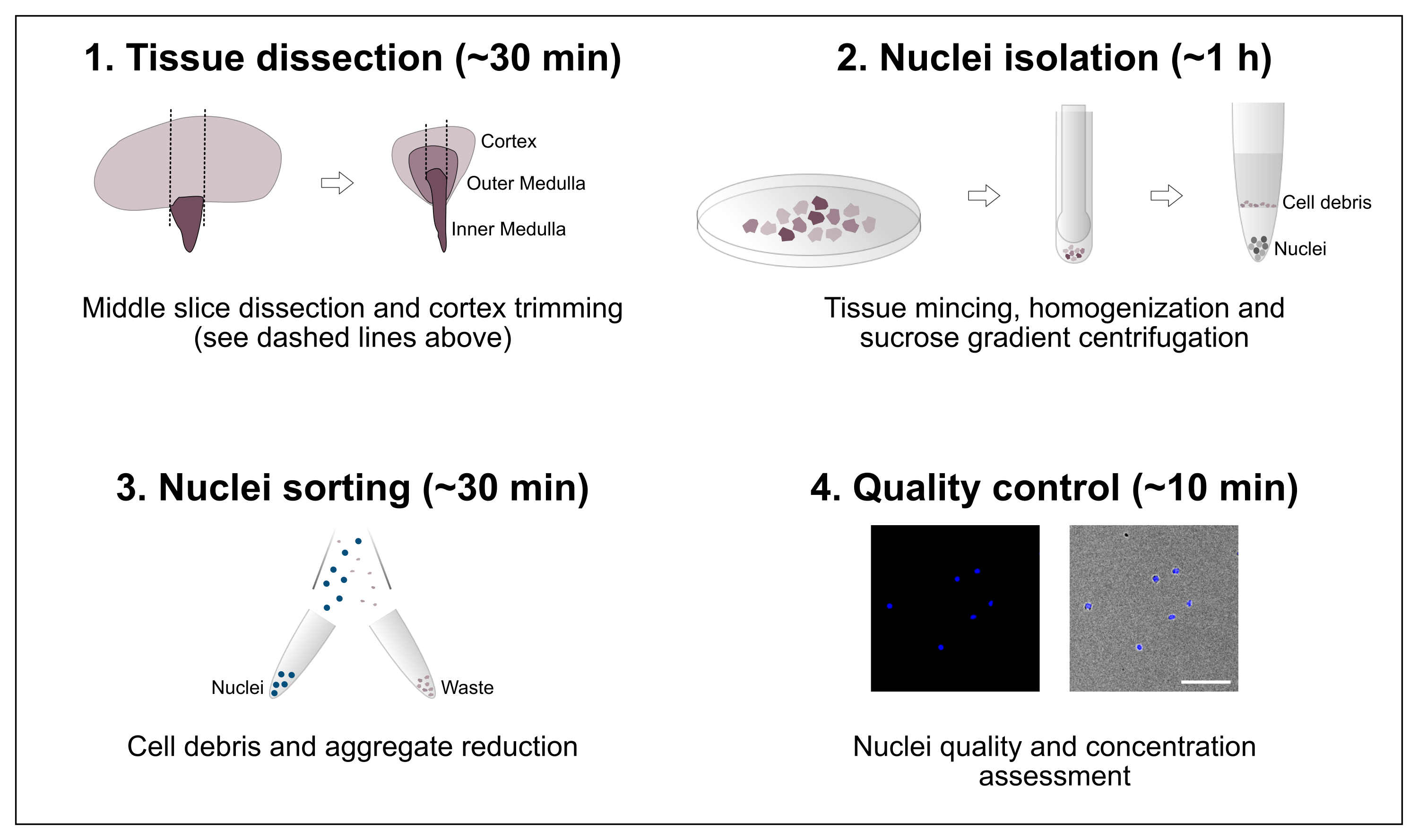
The protocol consists of four major steps that include tissue dissection followed by nuclei isolation, nuclei sorting and a final purity and concentration assessment. Scale bar = 100 µm.
This new approach opens the door to more accurate and representative RNA sequencing studies of the kidney. It is easy to implement, fast, and can be standardized across labs. By improving how researchers access and analyze cell-specific gene expression in the kidney, this technique could deepen our understanding of kidney diseases and support the development of better treatments.
Leiz J, Hinze C, Boltengagen A, Braeuning C, Kocks C, Rajewsky N, Schmidt-Ott KM. (2021) Nuclei isolation from adult mouse kidney for single-nucleus RNA-sequencing. JOVE 2021(175): e62901. [article]
Related Posts






Understanding how the kidney works at the level of individual cells is essential for studying health and disease. However, traditional methods for single-cell RNA sequencing (scRNA-seq) in kidney research come with major drawbacks. They typically require fresh tissue and can stress the cells during processing. This often leads to data that mostly reflects the outer part of the kidney, known as the cortex, while underrepresenting the medulla, the deeper inner regions.
To address this, researchers at the Universitätsmedizin Berlin have developed a new protocol that isolates nuclei instead of whole cells. This method works with frozen kidney tissue and is carried out entirely at cold temperatures, reducing stress on the cells and preserving RNA quality. More importantly, it allows scientists to capture a balanced view of all regions of the kidney, including the medulla, which plays a critical role in urine concentration and water balance.
Schematic overview of the workflow
The protocol consists of four major steps that include tissue dissection followed by nuclei isolation, nuclei sorting and a final purity and concentration assessment. Scale bar = 100 µm.
This new approach opens the door to more accurate and representative RNA sequencing studies of the kidney. It is easy to implement, fast, and can be standardized across labs. By improving how researchers access and analyze cell-specific gene expression in the kidney, this technique could deepen our understanding of kidney diseases and support the development of better treatments.
Leiz J, Hinze C, Boltengagen A, Braeuning C, Kocks C, Rajewsky N, Schmidt-Ott KM. (2021) Nuclei isolation from adult mouse kidney for single-nucleus RNA-sequencing. JOVE 2021(175): e62901. [article]
Related Posts
Recent Posts
 Scientists identify the neuroblastoma cells that signal poor outcomes
 GARAGE improves rare cell detection in single-cell RNA sequencing analysis using AI
 Machine learning reveals how RNA splicing is regulated in individual cell types
 Illumina releases SpliceAI2 to help advance rare disease research
 Postdoctoral Fellow – Long-Read RNA Methods Development for Neurodegeneration
 Novel biosensor uses DNA ‘barcodes’ to uncover disease signals in cells
RNA-Seq Products & Services


Stay Connected