

Researchers at the New York Genome Center have developed a new method called Paired-Damage-seq to study how DNA damage affects gene regulation at the single-cell level. DNA damage is a constant challenge for cells, arising from both environmental factors (like UV radiation) and internal processes (such as oxidative stress). Understanding where and how DNA damage occurs in different cell types is crucial for uncovering its role in diseases like cancer and neurodegeneration.
One of the biggest obstacles in studying DNA damage is the complexity of tissues, which contain many different cell types, each with distinct gene expression patterns. Additionally, DNA damage occurs randomly, making it difficult to pinpoint patterns of vulnerability. To address these challenges, the researchers developed Paired-Damage-seq, a technique that simultaneously analyzes oxidative and single-stranded DNA damage along with gene expression in individual cells.
Joint profiling of oxidative and single-stranded DNA damage with transcriptome in single cells
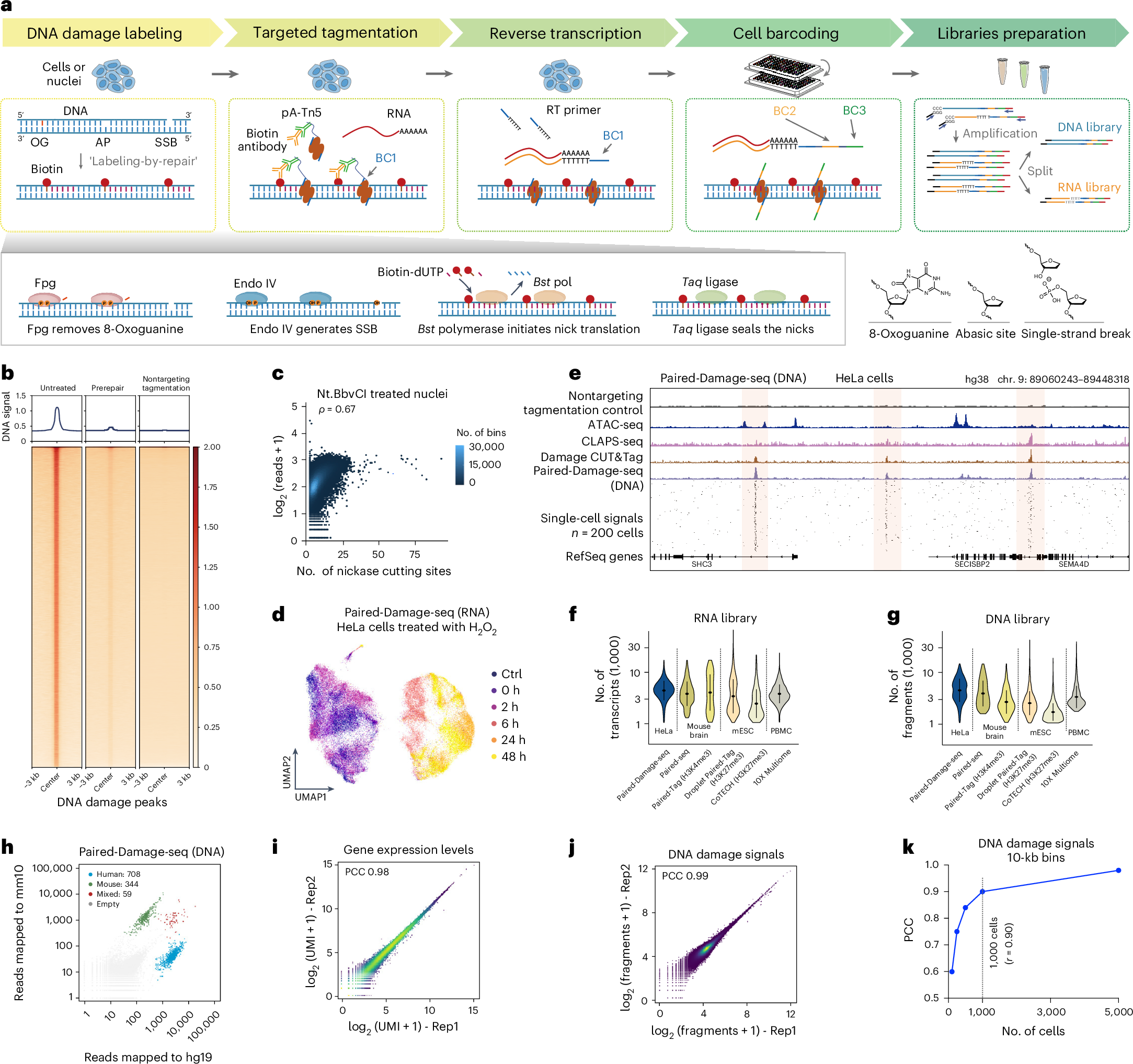
a, Schematic of the Paired-Damage-seq workflow with the ‘Labeling-by-repair’ strategy to label damage sites. OG, 8-oxoguanine; AP, apyrimidinic site; RT, reverse transcription. b, Heatmaps showing the reads densities on DNA damage hotspots for untreated, prerepair control and nontargeting tagmentation control profiles. c, Scatter plot showing the correlation between detected DNA damage reads densities with the numbers of Nt.BbvCI cutting sites per 10,000-bp nonoverlapping bins for nickase-treated nuclei. d, UMAP embedding showing single cells based on Paired-Damage-seq transcriptomic profiles. Each dot represents an individual cell and is colored according to the treatment conditions. Ctrl, control; 0, 2, 6, 24 and 48 h indicate the duration posttreatment in fresh medium. e, Representative genome browser view of pseudo-bulk and single-cell DNA damage signals of Paired-Damage-seq in HeLa cells. Bulk damage CUT&Tag, CLAPS-seq, ATAC-seq and nontargeting tagmentation control signals are also shown. f,g, Violin plots showing the unique number of transcripts (f) and unique number of DNA fragments per cell across datasets (g) generated with different single-cell multiomic assays. mESC, mouse embryonic stem cell; PBMC, peripheral blood mononuclear cell. For all box plots, hinges were drawn from the 25th to 75th percentiles, with the middle line denoting the median and whiskers denoting a maximum 2× the interquartile range. n = 64,794 (Paired-Damage-seq), 1,133 (Paired-seq RNA), 1,091 (Paired-seq DNA), 408 (Paired-Tag RNA), 468 (Paired-Tag DNA), 5,000 (Droplet Paired-Tag RNA), 4,455 (Droplet Paired-Tag DNA), 302 (CoTECH RNA), 3,031 (CoTECH DNA) and 11,614 (10X Multiome) cells. h, Scatter plots showing the fraction of DNA reads mapped to human and mouse reference genome for each cell barcode in the species-mixing experiment. i,j, Scatter plots showing the correlation of read counts (log2(fragments + 1)) from two technical replicates of Paired-Damage-seq RNA profiles (i) and DNA profiles (j). Rep1, replicate 1; Rep2, replicate 2. k, Line plot showing the correlation of DNA damage signals (at 10-kb nonoverlapping bins) between Bulk Paired-Damage-seq DNA profile and aggregated single-cell Paired-Damage-seq DNA signals from different numbers of cells.
The team tested this method in cultured HeLa cells and mouse brain tissue, demonstrating that certain areas of the genome are more prone to damage depending on the cell type. Interestingly, they found that oxidative DNA damage hotspots align with specific epigenetic markers, suggesting that chromatin structure influences genome stability. Moreover, the selective vulnerability of the genome in different cells can help predict which cell types are at higher risk for damage-related diseases.
This research represents a significant step forward in understanding the relationship between DNA damage and gene regulation. By using single-cell RNA sequencing, the scientists could link DNA damage patterns with gene expression changes, providing insights into how cells respond to genetic stress. Their findings may eventually lead to new ways to identify disease risks and develop targeted therapies based on individual genome vulnerabilities.
Availability – Custom scripts used for analyzing Paired-Damage-seq datasets are available from GitHub (https://github.com/czhulab/Paired-Damage-seq).
Bai D, Cao Z, Attada N, Song J, Zhu C. (2025) Single-cell parallel analysis of DNA damage and transcriptome reveals selective genome vulnerability. Nat Methods [Epub ahead of print]. [article]
Related Posts






Researchers at the New York Genome Center have developed a new method called Paired-Damage-seq to study how DNA damage affects gene regulation at the single-cell level. DNA damage is a constant challenge for cells, arising from both environmental factors (like UV radiation) and internal processes (such as oxidative stress). Understanding where and how DNA damage occurs in different cell types is crucial for uncovering its role in diseases like cancer and neurodegeneration.
One of the biggest obstacles in studying DNA damage is the complexity of tissues, which contain many different cell types, each with distinct gene expression patterns. Additionally, DNA damage occurs randomly, making it difficult to pinpoint patterns of vulnerability. To address these challenges, the researchers developed Paired-Damage-seq, a technique that simultaneously analyzes oxidative and single-stranded DNA damage along with gene expression in individual cells.
Joint profiling of oxidative and single-stranded DNA damage with transcriptome in single cells
a, Schematic of the Paired-Damage-seq workflow with the ‘Labeling-by-repair’ strategy to label damage sites. OG, 8-oxoguanine; AP, apyrimidinic site; RT, reverse transcription. b, Heatmaps showing the reads densities on DNA damage hotspots for untreated, prerepair control and nontargeting tagmentation control profiles. c, Scatter plot showing the correlation between detected DNA damage reads densities with the numbers of Nt.BbvCI cutting sites per 10,000-bp nonoverlapping bins for nickase-treated nuclei. d, UMAP embedding showing single cells based on Paired-Damage-seq transcriptomic profiles. Each dot represents an individual cell and is colored according to the treatment conditions. Ctrl, control; 0, 2, 6, 24 and 48 h indicate the duration posttreatment in fresh medium. e, Representative genome browser view of pseudo-bulk and single-cell DNA damage signals of Paired-Damage-seq in HeLa cells. Bulk damage CUT&Tag, CLAPS-seq, ATAC-seq and nontargeting tagmentation control signals are also shown. f,g, Violin plots showing the unique number of transcripts (f) and unique number of DNA fragments per cell across datasets (g) generated with different single-cell multiomic assays. mESC, mouse embryonic stem cell; PBMC, peripheral blood mononuclear cell. For all box plots, hinges were drawn from the 25th to 75th percentiles, with the middle line denoting the median and whiskers denoting a maximum 2× the interquartile range. n = 64,794 (Paired-Damage-seq), 1,133 (Paired-seq RNA), 1,091 (Paired-seq DNA), 408 (Paired-Tag RNA), 468 (Paired-Tag DNA), 5,000 (Droplet Paired-Tag RNA), 4,455 (Droplet Paired-Tag DNA), 302 (CoTECH RNA), 3,031 (CoTECH DNA) and 11,614 (10X Multiome) cells. h, Scatter plots showing the fraction of DNA reads mapped to human and mouse reference genome for each cell barcode in the species-mixing experiment. i,j, Scatter plots showing the correlation of read counts (log2(fragments + 1)) from two technical replicates of Paired-Damage-seq RNA profiles (i) and DNA profiles (j). Rep1, replicate 1; Rep2, replicate 2. k, Line plot showing the correlation of DNA damage signals (at 10-kb nonoverlapping bins) between Bulk Paired-Damage-seq DNA profile and aggregated single-cell Paired-Damage-seq DNA signals from different numbers of cells.
The team tested this method in cultured HeLa cells and mouse brain tissue, demonstrating that certain areas of the genome are more prone to damage depending on the cell type. Interestingly, they found that oxidative DNA damage hotspots align with specific epigenetic markers, suggesting that chromatin structure influences genome stability. Moreover, the selective vulnerability of the genome in different cells can help predict which cell types are at higher risk for damage-related diseases.
This research represents a significant step forward in understanding the relationship between DNA damage and gene regulation. By using single-cell RNA sequencing, the scientists could link DNA damage patterns with gene expression changes, providing insights into how cells respond to genetic stress. Their findings may eventually lead to new ways to identify disease risks and develop targeted therapies based on individual genome vulnerabilities.
Availability – Custom scripts used for analyzing Paired-Damage-seq datasets are available from GitHub (https://github.com/czhulab/Paired-Damage-seq).
Bai D, Cao Z, Attada N, Song J, Zhu C. (2025) Single-cell parallel analysis of DNA damage and transcriptome reveals selective genome vulnerability. Nat Methods [Epub ahead of print]. [article]
Related Posts
Recent Posts
 Machine learning reveals how RNA splicing is regulated in individual cell types
 Illumina releases SpliceAI2 to help advance rare disease research
 Postdoctoral Fellow – Long-Read RNA Methods Development for Neurodegeneration
 Novel biosensor uses DNA ‘barcodes’ to uncover disease signals in cells
 GP-DHT uses single-cell RNA sequencing to map gene regulatory networks
 Boehringer taps Envisagenics’ AI to mine RNA splicing for cancer targets
RNA-Seq Products & Services



Stay Connected