Researchers uncover an intriguing characteristic of pancreatic cancer that could detect – and potentially target – metastasis earlier.
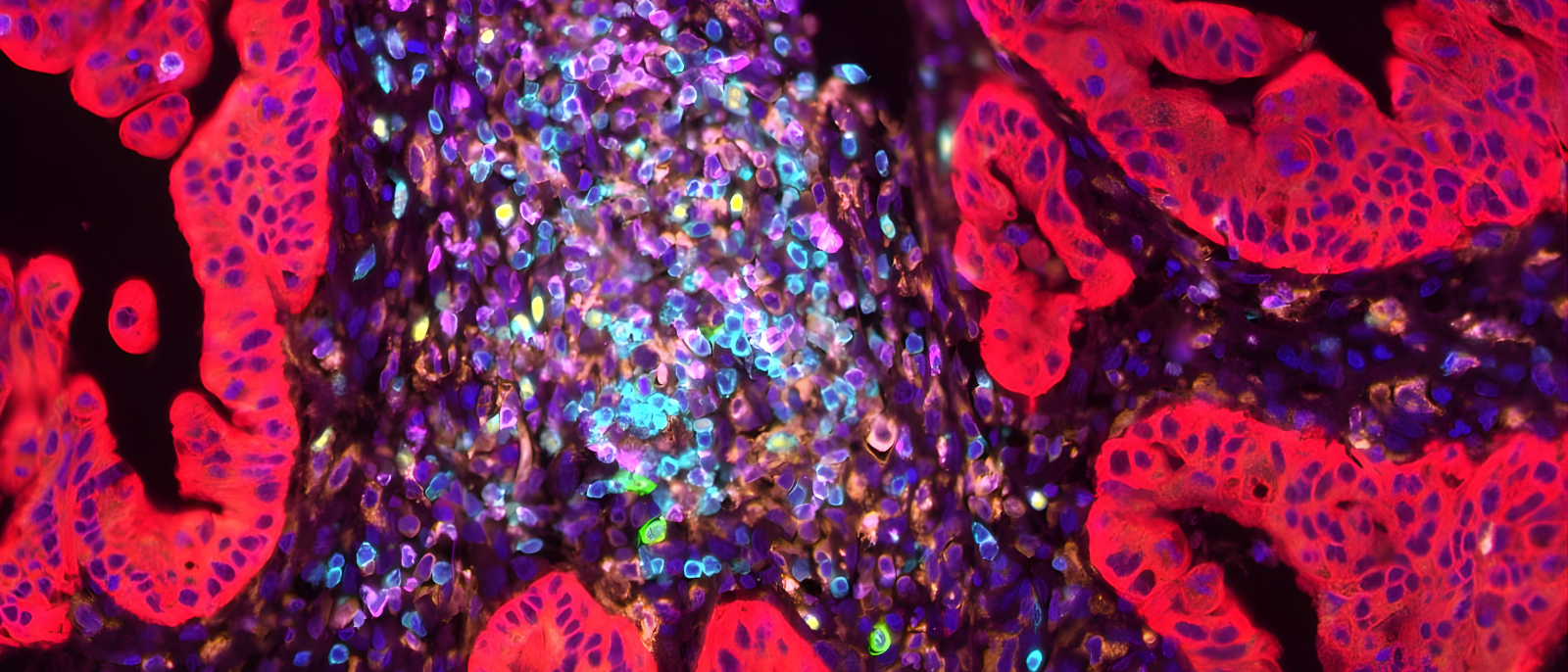
Even as they develop at their primary site, pancreatic cancer cells are already expressing the genes that will determine where they will metastasize, according to new findings from Columbia researchers. The work, published in Nature Genetics, reveals a new facet of cancer metastasis, and points toward novel strategies for anticipating and blocking this deadly phenomenon.
The project began a decade ago, when Gulam Manji, MD, PhD, co-leader of the Precision Oncology and Systems Biology research program at the Herbert Irving Comprehensive Cancer (HICCC), was struck by the dramatic differences in prognoses for pancreatic cancer patients. People whose tumors metastasized to their lungs alone did far better than those with liver metastases.
“It was very gratifying to see patients with lung only metastasis doing very well, yet at the same time really frustrating as an investigator, because there was biology within them that we didn’t really understand,” says Manji.
Clearing the hurdles
Columbia maintains a large repository of tumor specimens, but the experiment Manji had in mind required detailed clinical annotation of each sample. Enlisting the aid of collaborators, students, and postdocs, he had his team sift through several hundred cases, eventually finding 131 patients with isolated metastases of pancreatic tumors to either lung or liver.
The next challenge was analyzing the tumor specimens, which often contain mixed populations of cancer cells and normal cells of the immune system and adjacent tissues. To study them, the researchers developed new methods for single-nucleus RNA sequencing and whole genome sequencing, obtaining detailed data on individual cells’ gene expression patterns. Benjamin Izar, MD, PhD, member of the HICCC’s Tumor Biology and Microenvironment research program, led the computational biology side of the project, extracting and validating findings from the deluge of sequencing data.
A single-nucleus landscape of primary PDAC with initial liver or lung recurrence
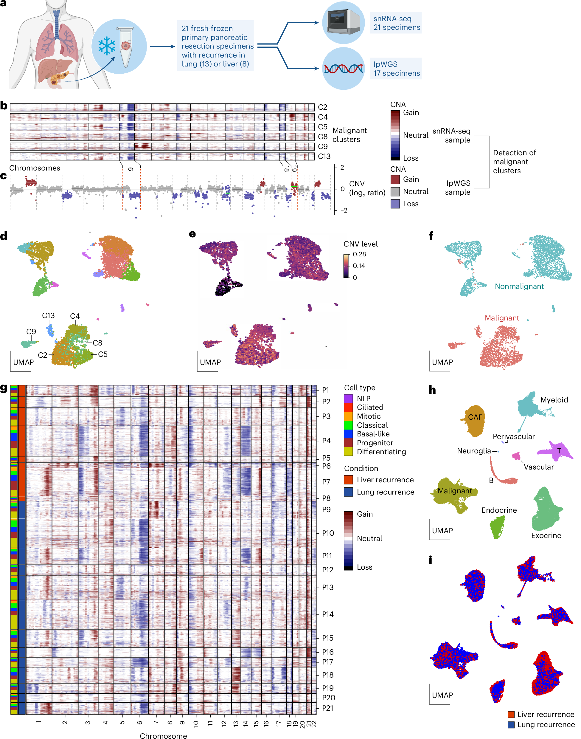
a, Experimental design of the CUIMC cohort. snRNA-seq was performed successfully on 21 (of 28) fresh-frozen specimens, and lpWGS data were available for 17 of these samples. b, CNVs inferred by inferCNV from snRNA-seq data for a representative patient, with rows representing malignant cells in six clusters and columns corresponding to genes across each chromosome. c, CNVs measured by ichorCNA from bulk lpWGS data of the patient in b, encompassing both malignant and nonmalignant cells. Dashed red lines indicate CNVs in corresponding chromosomes. d–f, UMAP visualizations of the clusters identified by Seurat (d), overall CNV burden (inferred by inferCNV) (e) and malignancy cell annotation (f) for the patient in b. The CNVs of the annotated cancer clusters in d are shown in b. g, CNVs inferred by inferCNV from snRNA-seq data for all malignant cells in the CUIMC cohort. Columns on the far left indicate malignant cell types and whether patients had lung or liver initial recurrence. P10 is the patient in b–f. h,i, snRNA-seq landscape of the CUIMC cohort (h) with initial recurrence sites (i). Labels P1–P21 represent chronological order and do not correspond to patient IDs (PNX)
Language lessons for prospective visitors
Using a suite of new technical and analytical advances, and by integrating several other data sets, Izar’s group stumbled over an interesting observation.
“Cancer cells from patient’s primary tumours that ended up relapsing in the liver or the lung early on adopt cellular states that resemble their ultimate target organ. In other words, they already spoke the right language to ultimately fit in into their future organ community,” says Izar.
Even at their primary site in the pancreas, the tumor cells are already impersonating cells of their destination organ, predisposing them to survive there. A key question was whether a specific change in the DNA caused this observation. The researchers applied machine-learning approaches and studied additional large patient data bases to try to find an answer.
“We could not identify a single genetic event that could explain tropism to either liver or lung. While we have to remain open to alternative explanations, so far, we could not find a genetic reason for this intriguing result,” Izar says.
The researchers also looked at cultured cells from other types of cancers and found conserved signatures suggesting the same sort of metastatic foresight.
“The key question now is what is the switch that takes these cells through these different programs?” says Manji.
While stressing that the results still need to be validated by other researchers and on more samples, Manji is excited about the effect they could have on patient care. In the near future, he hopes physicians will be able to refine surveillance strategies by predicting where a tumor is likely to metastasize. Looking further ahead, the gene expression profiles may point toward novel drug targets.
“Within the predictive signatures are likely embedded, determinants that allow these tumors to successfully establish a metastatic niche. Disrupting these key determinants is the next challenge which will likely produce a personalized drug for liver metastasis,” says Manji.
Source – Columbia University


Researchers uncover an intriguing characteristic of pancreatic cancer that could detect – and potentially target – metastasis earlier.
Even as they develop at their primary site, pancreatic cancer cells are already expressing the genes that will determine where they will metastasize, according to new findings from Columbia researchers. The work, published in Nature Genetics, reveals a new facet of cancer metastasis, and points toward novel strategies for anticipating and blocking this deadly phenomenon.
The project began a decade ago, when Gulam Manji, MD, PhD, co-leader of the Precision Oncology and Systems Biology research program at the Herbert Irving Comprehensive Cancer (HICCC), was struck by the dramatic differences in prognoses for pancreatic cancer patients. People whose tumors metastasized to their lungs alone did far better than those with liver metastases.
Clearing the hurdles
Columbia maintains a large repository of tumor specimens, but the experiment Manji had in mind required detailed clinical annotation of each sample. Enlisting the aid of collaborators, students, and postdocs, he had his team sift through several hundred cases, eventually finding 131 patients with isolated metastases of pancreatic tumors to either lung or liver.
The next challenge was analyzing the tumor specimens, which often contain mixed populations of cancer cells and normal cells of the immune system and adjacent tissues. To study them, the researchers developed new methods for single-nucleus RNA sequencing and whole genome sequencing, obtaining detailed data on individual cells’ gene expression patterns. Benjamin Izar, MD, PhD, member of the HICCC’s Tumor Biology and Microenvironment research program, led the computational biology side of the project, extracting and validating findings from the deluge of sequencing data.
A single-nucleus landscape of primary PDAC with initial liver or lung recurrence
a, Experimental design of the CUIMC cohort. snRNA-seq was performed successfully on 21 (of 28) fresh-frozen specimens, and lpWGS data were available for 17 of these samples. b, CNVs inferred by inferCNV from snRNA-seq data for a representative patient, with rows representing malignant cells in six clusters and columns corresponding to genes across each chromosome. c, CNVs measured by ichorCNA from bulk lpWGS data of the patient in b, encompassing both malignant and nonmalignant cells. Dashed red lines indicate CNVs in corresponding chromosomes. d–f, UMAP visualizations of the clusters identified by Seurat (d), overall CNV burden (inferred by inferCNV) (e) and malignancy cell annotation (f) for the patient in b. The CNVs of the annotated cancer clusters in d are shown in b. g, CNVs inferred by inferCNV from snRNA-seq data for all malignant cells in the CUIMC cohort. Columns on the far left indicate malignant cell types and whether patients had lung or liver initial recurrence. P10 is the patient in b–f. h,i, snRNA-seq landscape of the CUIMC cohort (h) with initial recurrence sites (i). Labels P1–P21 represent chronological order and do not correspond to patient IDs (PNX)
Language lessons for prospective visitors
Using a suite of new technical and analytical advances, and by integrating several other data sets, Izar’s group stumbled over an interesting observation.
Even at their primary site in the pancreas, the tumor cells are already impersonating cells of their destination organ, predisposing them to survive there. A key question was whether a specific change in the DNA caused this observation. The researchers applied machine-learning approaches and studied additional large patient data bases to try to find an answer.
The researchers also looked at cultured cells from other types of cancers and found conserved signatures suggesting the same sort of metastatic foresight.
While stressing that the results still need to be validated by other researchers and on more samples, Manji is excited about the effect they could have on patient care. In the near future, he hopes physicians will be able to refine surveillance strategies by predicting where a tumor is likely to metastasize. Looking further ahead, the gene expression profiles may point toward novel drug targets.
Source – Columbia University
Chalabi Hajkarim M, May M, Amin A D, Jamison J, Tagore S, D’Souza E, Caprio L, Walsh Z H, Fan J L, Zhang M, Shah P, Abuzaid S, Michel A, Dajiang S, Wong W, Shaikh N, Ramaradj P, Bakir B, Kluger M D, Chabot J, Azizi E, Hibshoosh H, Rustgi A K, Raufi A G, Manji G A, Izar B. (2025) Cellular states associated with metastatic organotropism and survival in patients with pancreatic ductal adenocarcinoma. Nature Genetics 57(11): 2728–2742. [article]
Related Posts
Novel biosensor uses DNA ‘barcodes’ to uncover disease signals in cells
CellART extracts single-cell information from high-resolution spatial transcriptomics
Cell2space restores spatial context to single-cell RNA sequencing data
Urine test may improve bladder cancer diagnosis and guide treatment decisions
DeCoRE – a computational method to resolve RNA structural heterogeneity from RNA structure probing data by direct RNA sequencing
Tracing 700 million years of cell evolution
How should biological replicates be handled in long-read RNA sequencing?
RNA-Seq reveals a new way the developing heart controls its growth
The Human RNome Project Consortium seeks to publish first-ever complete sequence of the body’s RNA
scFlowReport brings multiple single-cell RNA sequencing analyses into one workflow
Long-read RNA sequencing reveals how neurons choose alternative RNA endings
DeOPUS improves cell-type estimates from bulk RNA sequencing
ResolVI improves accuracy in spatial transcriptomics analysis
RNA sequencing reveals transcriptional allele differences in prostate cancer stem cells
Unmasking “zombie cells” in aging tissue with an AI-powered barcode
RNA variant imaging for cancer diagnosis
Autism-risk mutations reveal two opposing patterns of brain gene activity
A real-data approach to evaluating RNA transcript analysis methods
Detection and sequencing of Ap2N-capped RNAs in human cells
Single-cell RNA sequencing provides a closer look at the Aedes aegypti midgut
Stay Connected
Submit a Post to the Blog
Recent Posts
Subscribe to the RNA-Seq Blog
RNA-Seq Products & Services