


Understanding how genes influence cancer growth and spread requires tools that can connect genetic changes to what is happening inside tissues. While CRISPR screening has become a powerful way to test the function of thousands of genes at once, researchers have faced challenges when trying to examine these effects within the natural architecture of tissues. Existing approaches have generally been limited to smaller gene libraries and have focused primarily on protein-coding genes.
Researchers from the Department of Biomedical Engineering at Yale University have developed a new technology called Perturb-DBiT that significantly expands the scope of spatial CRISPR screening. The method allows scientists to simultaneously measure spatial RNA expression patterns and identify CRISPR guide RNAs within the same tissue section. This provides a detailed view of how genetic perturbations affect cells in their native environment.
Perturb-DBiT combines large-scale CRISPR screening with spatial transcriptomics and total RNA profiling. Rather than examining only protein-coding messenger RNAs, the technique captures a broad range of RNA molecules, including long noncoding RNAs, microRNAs, transfer RNAs, and messenger RNAs. This comprehensive approach helps researchers understand how different layers of gene regulation contribute to disease processes.
Perturb-DBiT overview, technical performance and demonstration of robust performance revealing tumor clonal heterogeneity in medium-sized CRISPR screening libraries
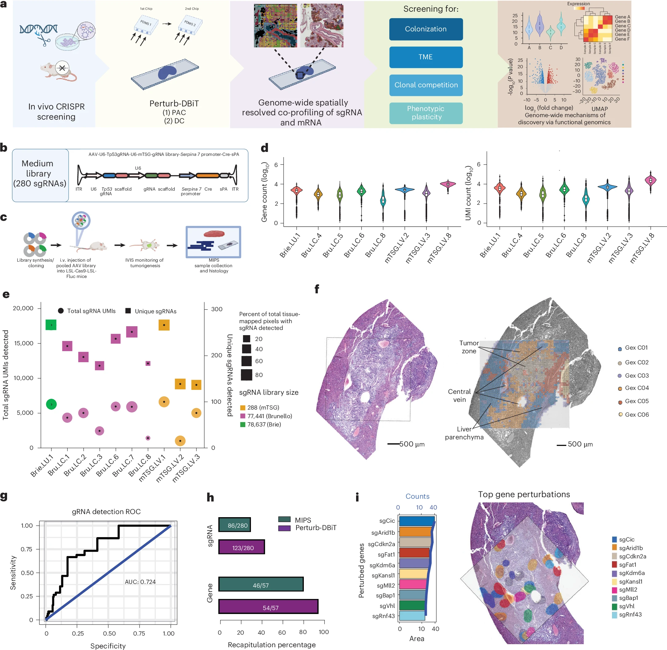
a, Schematic of Perturb-DBiT and its applications. b, Schematic of the AAV-CRISPR vector containing two sgRNA-expressing cassettes used for Perturb-DBiT with the medium (288-sgRNA) library; ITR, inverted terminal repeat; sPA, synthetic polyadenylation signal. c, Schematic of direct in vivo AAV-CRISPR liver screen design; i.v., intravenous. d, Violin plots showing the distribution of log10-transformed detected gene counts (left) and UMI counts (right) per spatial capture spot across eight Perturb-DBiT samples. The unit of study is the individual spatial spot, which represents a technical measurement nested within the biological samples. Within each violin plot, the central white box indicates the interquartile range (25th to 75th percentiles), the internal horizontal line denotes the median, and the whiskers extend to the maximum and minimum values within 1.5× interquartile range. Points beyond the whiskers represent outlier spatial spots. The precise number (n) of spatial spots analyzed for each sample is n = 1,160 (Brie.LU.1), n = 2,115 (Bru.LC.4), n = 2,170 (Bru.LC.5), n = 2,173 (Bru.LC.6), n = 6,398 (Bru.LC.8), n = 2,148 (mTSG.LV.2), n = 2,360 (mTSG.LV.3) and n = 2,256 (PD.Bru.LC.6). The eight samples correspond to tumors derived from multiple CRISPR screening mouse models; in some cases, multiple tumors or sections originate from the same mouse. Because these data evaluate descriptive transcript capture distributions rather than hypothesis testing, no statistical comparisons or specific control groups are evaluated. Detailed sample information, including mouse origin and tumor model, is provided in Extended Data Fig. 1e. e, Bubble plot depicting Perturb-DBiT sgRNA capture diversity. f, Detection of a medium-sized guide library (288 sgRNAs) in a mouse model of autochthonous liver cancer. Left, liver tissue stained with H&E, demonstrating the ROI used for Perturb-DBiT. Right, spatial clustering of gene expression data overlaid on a brightfield image, labeled with pathologist annotations. The representative experiment shown here was repeated independently with similar results. g, ROC curve comparing sgRNA detection by 50-μm Perturb-DBiT versus the traditional pooled sequencing method (MIPS). h, Recapitulation analysis comparing sgRNA capture accuracy between Perturb-DBiT and the traditional pooled sequencing method (MIPS). i, Left, bar plot of the top ten gene perturbations detected by 50-μm Perturb-DBiT. The area and total counts of the gene perturbations are presented by bars and the blue line, respectively. Right, spatial visualization of the top gene perturbations, depicted by color-coded 2D density maps on top of an autochthonous liver tumor ROI.
To demonstrate the technology, the research team applied an enormous CRISPR library containing more than 80,000 single guide RNAs in a human cancer metastasis model. By analyzing tumor colonies across multiple tissue sections, they were able to connect specific genetic perturbations to changes in RNA expression and tumor behavior.
The findings revealed that several types of RNA regulation influence cancer progression. Changes affecting long noncoding RNAs, microRNA-mRNA interactions, and amino acid-specific transfer RNAs were associated with tumor migration and growth. These results highlight the importance of RNA molecules that were previously difficult to study in large-scale spatial screening experiments.
The researchers also used computational methods to reconstruct transcriptional pseudotime trajectories, allowing them to track how tumor cell populations evolved over time. This analysis showed how different genetic perturbations altered clonal dynamics and cooperative behaviors among cancer cells. Such insights may help scientists better understand why some cancer cells become more aggressive or resistant to treatment.
In a separate immune-competent mouse model, Perturb-DBiT was used to examine interactions between tumors and the immune system. The technology revealed distinct combinations of genetic perturbations that influenced immune cell infiltration and immune suppression within the tumor microenvironment. These observations could help identify new therapeutic strategies that improve anti-tumor immune responses.
One of the most significant advantages of Perturb-DBiT is its ability to capture both spatial context and comprehensive RNA information simultaneously. Cancer cells do not exist in isolation, they interact continuously with neighboring cells, immune cells, and surrounding tissues. By preserving this spatial information, researchers can gain a more accurate understanding of how genetic changes shape disease progression.
The development of Perturb-DBiT represents an important step forward for functional genomics, cancer biology, and RNA sequencing research. By enabling large-scale, spatially resolved CRISPR screening while capturing the full complexity of RNA regulation, the method provides a powerful new framework for investigating tumor growth, metastasis, immune interactions, and other biological processes in complex tissues.
Availability – All custom code, scripts and analytical pipelines used to process the Perturb-DBiT data are available on GitHub at https://github.com/abaysoy/Perturb_DBiT.
Baysoy A, Tian X, Renauer P, Zhang F, Bai Z, Shi H, Yang M, Zhang D, Liu M, Li H, Tao B, Enninful A, Lu Y, Gao F, Wang G, Zhang W, Tran T, Patterson NH, Sheng J, Bao S, Dong C, Xin S, Chen B, Zhong M, Rankin S, Guy C, Wang Y, Connelly JP, Pruett-Miller SM, Wang D, Xu M, Gerstein MB, Chi H, Chen S, Fan R. (2026) Large-scale, spatially resolved panoramic CRISPR screening in native tissue environments using Perturb-DBiT. Nature Biotechnology [Epub ahead of print]. [article]
Related Posts






Understanding how genes influence cancer growth and spread requires tools that can connect genetic changes to what is happening inside tissues. While CRISPR screening has become a powerful way to test the function of thousands of genes at once, researchers have faced challenges when trying to examine these effects within the natural architecture of tissues. Existing approaches have generally been limited to smaller gene libraries and have focused primarily on protein-coding genes.
Researchers from the Department of Biomedical Engineering at Yale University have developed a new technology called Perturb-DBiT that significantly expands the scope of spatial CRISPR screening. The method allows scientists to simultaneously measure spatial RNA expression patterns and identify CRISPR guide RNAs within the same tissue section. This provides a detailed view of how genetic perturbations affect cells in their native environment.
Perturb-DBiT combines large-scale CRISPR screening with spatial transcriptomics and total RNA profiling. Rather than examining only protein-coding messenger RNAs, the technique captures a broad range of RNA molecules, including long noncoding RNAs, microRNAs, transfer RNAs, and messenger RNAs. This comprehensive approach helps researchers understand how different layers of gene regulation contribute to disease processes.
Perturb-DBiT overview, technical performance and demonstration of robust performance revealing tumor clonal heterogeneity in medium-sized CRISPR screening libraries
a, Schematic of Perturb-DBiT and its applications. b, Schematic of the AAV-CRISPR vector containing two sgRNA-expressing cassettes used for Perturb-DBiT with the medium (288-sgRNA) library; ITR, inverted terminal repeat; sPA, synthetic polyadenylation signal. c, Schematic of direct in vivo AAV-CRISPR liver screen design; i.v., intravenous. d, Violin plots showing the distribution of log10-transformed detected gene counts (left) and UMI counts (right) per spatial capture spot across eight Perturb-DBiT samples. The unit of study is the individual spatial spot, which represents a technical measurement nested within the biological samples. Within each violin plot, the central white box indicates the interquartile range (25th to 75th percentiles), the internal horizontal line denotes the median, and the whiskers extend to the maximum and minimum values within 1.5× interquartile range. Points beyond the whiskers represent outlier spatial spots. The precise number (n) of spatial spots analyzed for each sample is n = 1,160 (Brie.LU.1), n = 2,115 (Bru.LC.4), n = 2,170 (Bru.LC.5), n = 2,173 (Bru.LC.6), n = 6,398 (Bru.LC.8), n = 2,148 (mTSG.LV.2), n = 2,360 (mTSG.LV.3) and n = 2,256 (PD.Bru.LC.6). The eight samples correspond to tumors derived from multiple CRISPR screening mouse models; in some cases, multiple tumors or sections originate from the same mouse. Because these data evaluate descriptive transcript capture distributions rather than hypothesis testing, no statistical comparisons or specific control groups are evaluated. Detailed sample information, including mouse origin and tumor model, is provided in Extended Data Fig. 1e. e, Bubble plot depicting Perturb-DBiT sgRNA capture diversity. f, Detection of a medium-sized guide library (288 sgRNAs) in a mouse model of autochthonous liver cancer. Left, liver tissue stained with H&E, demonstrating the ROI used for Perturb-DBiT. Right, spatial clustering of gene expression data overlaid on a brightfield image, labeled with pathologist annotations. The representative experiment shown here was repeated independently with similar results. g, ROC curve comparing sgRNA detection by 50-μm Perturb-DBiT versus the traditional pooled sequencing method (MIPS). h, Recapitulation analysis comparing sgRNA capture accuracy between Perturb-DBiT and the traditional pooled sequencing method (MIPS). i, Left, bar plot of the top ten gene perturbations detected by 50-μm Perturb-DBiT. The area and total counts of the gene perturbations are presented by bars and the blue line, respectively. Right, spatial visualization of the top gene perturbations, depicted by color-coded 2D density maps on top of an autochthonous liver tumor ROI.
To demonstrate the technology, the research team applied an enormous CRISPR library containing more than 80,000 single guide RNAs in a human cancer metastasis model. By analyzing tumor colonies across multiple tissue sections, they were able to connect specific genetic perturbations to changes in RNA expression and tumor behavior.
The findings revealed that several types of RNA regulation influence cancer progression. Changes affecting long noncoding RNAs, microRNA-mRNA interactions, and amino acid-specific transfer RNAs were associated with tumor migration and growth. These results highlight the importance of RNA molecules that were previously difficult to study in large-scale spatial screening experiments.
The researchers also used computational methods to reconstruct transcriptional pseudotime trajectories, allowing them to track how tumor cell populations evolved over time. This analysis showed how different genetic perturbations altered clonal dynamics and cooperative behaviors among cancer cells. Such insights may help scientists better understand why some cancer cells become more aggressive or resistant to treatment.
In a separate immune-competent mouse model, Perturb-DBiT was used to examine interactions between tumors and the immune system. The technology revealed distinct combinations of genetic perturbations that influenced immune cell infiltration and immune suppression within the tumor microenvironment. These observations could help identify new therapeutic strategies that improve anti-tumor immune responses.
One of the most significant advantages of Perturb-DBiT is its ability to capture both spatial context and comprehensive RNA information simultaneously. Cancer cells do not exist in isolation, they interact continuously with neighboring cells, immune cells, and surrounding tissues. By preserving this spatial information, researchers can gain a more accurate understanding of how genetic changes shape disease progression.
The development of Perturb-DBiT represents an important step forward for functional genomics, cancer biology, and RNA sequencing research. By enabling large-scale, spatially resolved CRISPR screening while capturing the full complexity of RNA regulation, the method provides a powerful new framework for investigating tumor growth, metastasis, immune interactions, and other biological processes in complex tissues.
Availability – All custom code, scripts and analytical pipelines used to process the Perturb-DBiT data are available on GitHub at https://github.com/abaysoy/Perturb_DBiT.
Baysoy A, Tian X, Renauer P, Zhang F, Bai Z, Shi H, Yang M, Zhang D, Liu M, Li H, Tao B, Enninful A, Lu Y, Gao F, Wang G, Zhang W, Tran T, Patterson NH, Sheng J, Bao S, Dong C, Xin S, Chen B, Zhong M, Rankin S, Guy C, Wang Y, Connelly JP, Pruett-Miller SM, Wang D, Xu M, Gerstein MB, Chi H, Chen S, Fan R. (2026) Large-scale, spatially resolved panoramic CRISPR screening in native tissue environments using Perturb-DBiT. Nature Biotechnology [Epub ahead of print]. [article]
Related Posts
Recent Posts
 Machine learning reveals how RNA splicing is regulated in individual cell types
 Illumina releases SpliceAI2 to help advance rare disease research
 Postdoctoral Fellow – Long-Read RNA Methods Development for Neurodegeneration
 Novel biosensor uses DNA ‘barcodes’ to uncover disease signals in cells
 GP-DHT uses single-cell RNA sequencing to map gene regulatory networks
 Boehringer taps Envisagenics’ AI to mine RNA splicing for cancer targets
RNA-Seq Products & Services


Stay Connected