
A two-stage program where septoclasts initially degrade cartilage, followed by bone marrow stromal cells (BMSCs) developing the bone marrow
Bone marrow is the spongy tissue located within the hollow center of bones, serving as the primary site for the continuous production of red blood cells, platelets, and white blood cells. Despite its physiological importance, the developmental mechanism by which this soft tissue is formed within the rigid confines of hard bone has remained largely unknown.
A new study published on April 14, 2026, in Nature Communications now clarifies this development, identifying a two-step process where cell populations act as “organizers” to build the bone marrow environment. Specifically, specialized cells that degrade cartilage, called septoclasts, create a space inside the developing bone so that bone marrow stromal cells can sustain and grow the marrow environment.
This investigation began with the hypothesis that bone marrow development is coordinated by so-called “organizer cells,” a group of specialized cells that coordinate organ formation.
“Just as lymphoid tissue inducer cells initiate lymph node formation, we hypothesized that a distinct cell population may similarly serve as an organizer to trigger the formation of the bone marrow cavity,” explained Professor Shinichiro Sawa from Kyushu University’s Medical Institute of Bioregulation.
A primary candidate for this organizing signal is the cytokine RANKL, a protein already known for its role in bone remodeling. Previous evidence showed that mice lacking RANKL fail to develop bone-resorbing osteoclasts and do not form a proper bone marrow cavity. This suggested that RANKL-producing cells might serve as a previously unrecognized organizing niche that facilitates bone marrow formation.
The researchers put this hypothesis to the test by creating new RANKL reporter mice, which enable visualization of the RANKL gene throughout fetal development. Studying the early development of the tibia, the team observed the first RANKL-producing cells emerging in the central cortical region of the bone, which then invaded the cartilage. These potent signaling cells were found concentrated at the boundary where cartilage meets developing bone, positioned in close proximity to osteocytes.
Using single-cell RNA sequencing, the team characterized these early RANKL-producing cells as septoclasts. These specialized phagocytes produce enzymes that degrade the cartilaginous matrix. The findings suggest that fetal septoclasts coordinate the replacement of cartilage with bone marrow by clearing the extracellular matrix and inducing osteoclasts.
Developmental process of bone and bone marrow
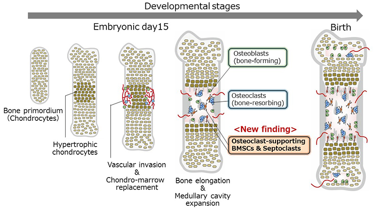
An illustration of bone and bone marrow development. During the process, osteoclasts (cells in blue) expand the marrow cavity by absorbing and removing unnecessary bone. In this study, the team found that in early bone development septoclasts (cells in red) and bone marrow stromal cells (BMSCs: cells in green) play a two-step role to build bone marrow.
As development progresses, the responsibility for RANKL production shifts to a different cell population. The study found that at later stages, Leptin receptor (LepR)-expressing bone marrow stromal cells (BMSCs) become the primary producers of RANKL.
Taken together, the research demonstrates that bone marrow cavity formation is a hardwired developmental process supported by a “relay” between two distinct cell populations: septoclasts at the early stage and LepR+ BMSCs at the later stage.
The team further leveraged the RANKL-reporter mice to identify the physiological sources of RANKL during bone repair. Following a fracture, they observed the emergence of RANKL-producing cells that correspond precisely to septoclasts and LepR+ BMSCs. This discovery suggests that fracture repair effectively reactivates the specialized cellular program originally deployed during fetal bone marrow formation.
These findings have significant implications for treating bone diseases such as osteoporosis and rheumatoid arthritis, in which excessive bone destruction occurs. By identifying the cells supporting osteoclasts, the study reveals a new therapeutic target. Focusing on these organizer cells may enable more precise and sustained control of pathological bone resorption.
Source – Kyushu University


A two-stage program where septoclasts initially degrade cartilage, followed by bone marrow stromal cells (BMSCs) developing the bone marrow
Bone marrow is the spongy tissue located within the hollow center of bones, serving as the primary site for the continuous production of red blood cells, platelets, and white blood cells. Despite its physiological importance, the developmental mechanism by which this soft tissue is formed within the rigid confines of hard bone has remained largely unknown.
A new study published on April 14, 2026, in Nature Communications now clarifies this development, identifying a two-step process where cell populations act as “organizers” to build the bone marrow environment. Specifically, specialized cells that degrade cartilage, called septoclasts, create a space inside the developing bone so that bone marrow stromal cells can sustain and grow the marrow environment.
This investigation began with the hypothesis that bone marrow development is coordinated by so-called “organizer cells,” a group of specialized cells that coordinate organ formation.
A primary candidate for this organizing signal is the cytokine RANKL, a protein already known for its role in bone remodeling. Previous evidence showed that mice lacking RANKL fail to develop bone-resorbing osteoclasts and do not form a proper bone marrow cavity. This suggested that RANKL-producing cells might serve as a previously unrecognized organizing niche that facilitates bone marrow formation.
The researchers put this hypothesis to the test by creating new RANKL reporter mice, which enable visualization of the RANKL gene throughout fetal development. Studying the early development of the tibia, the team observed the first RANKL-producing cells emerging in the central cortical region of the bone, which then invaded the cartilage. These potent signaling cells were found concentrated at the boundary where cartilage meets developing bone, positioned in close proximity to osteocytes.
Using single-cell RNA sequencing, the team characterized these early RANKL-producing cells as septoclasts. These specialized phagocytes produce enzymes that degrade the cartilaginous matrix. The findings suggest that fetal septoclasts coordinate the replacement of cartilage with bone marrow by clearing the extracellular matrix and inducing osteoclasts.
Developmental process of bone and bone marrow
An illustration of bone and bone marrow development. During the process, osteoclasts (cells in blue) expand the marrow cavity by absorbing and removing unnecessary bone. In this study, the team found that in early bone development septoclasts (cells in red) and bone marrow stromal cells (BMSCs: cells in green) play a two-step role to build bone marrow.
As development progresses, the responsibility for RANKL production shifts to a different cell population. The study found that at later stages, Leptin receptor (LepR)-expressing bone marrow stromal cells (BMSCs) become the primary producers of RANKL.
Taken together, the research demonstrates that bone marrow cavity formation is a hardwired developmental process supported by a “relay” between two distinct cell populations: septoclasts at the early stage and LepR+ BMSCs at the later stage.
The team further leveraged the RANKL-reporter mice to identify the physiological sources of RANKL during bone repair. Following a fracture, they observed the emergence of RANKL-producing cells that correspond precisely to septoclasts and LepR+ BMSCs. This discovery suggests that fracture repair effectively reactivates the specialized cellular program originally deployed during fetal bone marrow formation.
These findings have significant implications for treating bone diseases such as osteoporosis and rheumatoid arthritis, in which excessive bone destruction occurs. By identifying the cells supporting osteoclasts, the study reveals a new therapeutic target. Focusing on these organizer cells may enable more precise and sustained control of pathological bone resorption.
Source – Kyushu University
Sumiya E, Saeki K, Nakano K, Kikutake C, Kurisaki N, Nakaima N, Kurumata-Shigeto M, Kitada Y, Morioka Y, Go Y, Suyama M, Yoshimura Y, Goto M, Ito M, Nakayama M, Akiyama H, Peduto L, Okamura T, Matsushita Y, Sawa S. (2026) Medullary cavity expansion is mediated by distinct cell populations during fetal bone development. Nature Communications [Epub ahead of print]. [article]
Related Posts
RNAmel simplifies RNA sequencing data analysis with a reproducible, integrated platform
Heart failure reshapes bone marrow to create inflammation-related immune memory
Overlooked DNA regions surrounding ‘microexons’ hold missing pieces of autism’s genetic puzzle
RiboBA improves detection of hidden protein-coding regions using ribosome profiling
An anti-senescence gene may fight spinal disc deterioration
Scientists identify the neuroblastoma cells that signal poor outcomes
Novel biosensor uses DNA ‘barcodes’ to uncover disease signals in cells
CellART extracts single-cell information from high-resolution spatial transcriptomics
Cell2space restores spatial context to single-cell RNA sequencing data
Urine test may improve bladder cancer diagnosis and guide treatment decisions
DeCoRE – a computational method to resolve RNA structural heterogeneity from RNA structure probing data by direct RNA sequencing
Tracing 700 million years of cell evolution
How should biological replicates be handled in long-read RNA sequencing?
RNA-Seq reveals a new way the developing heart controls its growth
The Human RNome Project Consortium seeks to publish first-ever complete sequence of the body’s RNA
scFlowReport brings multiple single-cell RNA sequencing analyses into one workflow
Long-read RNA sequencing reveals how neurons choose alternative RNA endings
DeOPUS improves cell-type estimates from bulk RNA sequencing
ResolVI improves accuracy in spatial transcriptomics analysis
RNA sequencing reveals transcriptional allele differences in prostate cancer stem cells
Stay Connected
Submit a Post to the Blog
Recent Posts
Subscribe to the RNA-Seq Blog
RNA-Seq Products & Services