

Understanding the smallest changes in RNA is key to uncovering the root causes of many diseases. However, detecting these subtle mutations and chemical modifications has long been a challenge due to the limitations of traditional techniques. Now, scientists at the University of Cambridge have introduced an innovative approach called RNA-SCAN that offers a precise, simple, and scalable way to analyze RNA at the single-nucleotide level.
RNA mutations and modifications are often implicated in serious conditions, ranging from cancer to infections. However, existing detection methods are complex, error-prone, and require heavy data processing, which limits their clinical and research use. The RNA-SCAN system solves these challenges by combining programmable nanotechnology with solid-state nanopore sensing, providing a powerful new tool for RNA analysis.
At the heart of RNA-SCAN is a “nanolatch,” a short DNA strand specifically designed to bind to a target RNA sequence. The system measures how well this latch binds to the RNA as it passes through a nanopore, a microscopic hole that allows researchers to monitor individual molecules in real time. When there’s a mismatch or chemical modification in the RNA, the binding pattern changes. These variations are detected through shifts in signal intensity and frequency, which are recorded and analyzed at the single-molecule level.
RNA-SCAN: a nanolatch-nanopore sensor for nucleic acid characterization
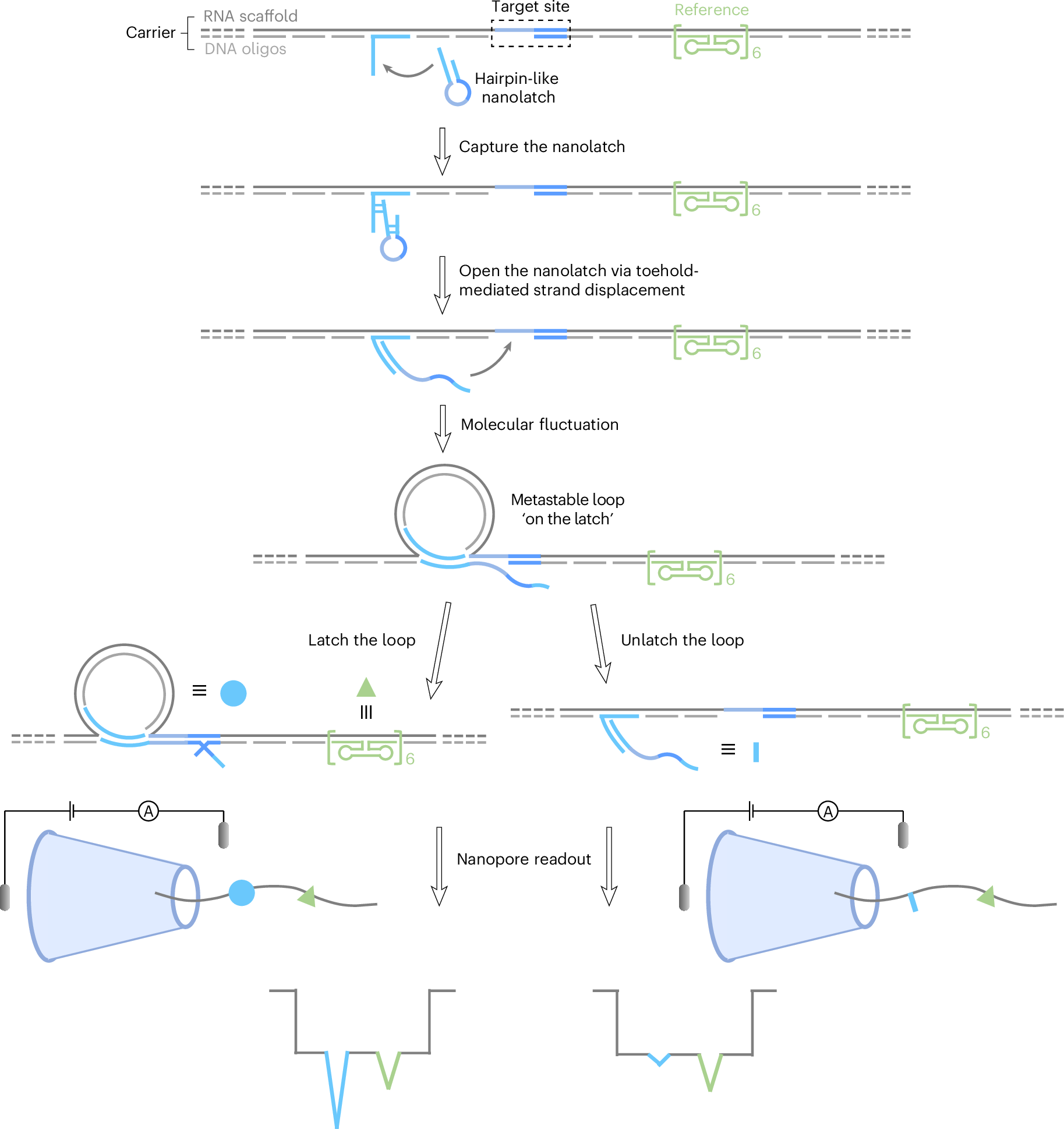
The RNA-SCAN mechanism proceeds as follows (where the complementary strand regions indicated by matching colours). A hairpin-like nanolatch is first captured and opened by an overhang on the RNA/DNA carrier (self-assembled from a long RNA scaffold and short DNA oligos) through toehold-mediated strand displacement, exposing its target site-complementary segment. Molecular fluctuations in solution then bring the nanolatch’s free end into proximity to the target site, forming a metastable loop structure referred to as the ‘on the latch’ state, a configuration that is formed but not yet fastened. The subsequent stability of the loop depends on base-pairing strength between the nanolatch and target site, resulting in either a ‘latched’ state (where strong complementarity fastens the loop) or an ‘unlatched’ state (where weak binding leads to loop opening). During nanopore detection, the translocation of the carrier generates a characteristic baseline current drop. A six-dumbbell structure (marked in green) positioned next to the target site consistently produces a small spike of approximately half the amplitude of the first-level current drop generated by the carrier alone, serving as a reference. In comparison, a latched loop typically generates a spike larger than the reference spike, approaching the magnitude of the first-level current drop, while an unlatched loop yields minimal current fluctuation beyond the plateau.
To validate their method, the researchers used RNA from two closely related bacterial species, Escherichia coli and Salmonella spp., whose genetic material differs at only a few nucleotides. RNA-SCAN successfully identified these small differences and even distinguished chemical modifications such as m5C1407, a common epigenetic mark in bacterial RNA. Impressively, the system could also quantify different Salmonella strains in mixed samples, showcasing its potential for infection diagnostics and microbial surveillance.
What makes RNA-SCAN particularly promising is its simplicity. It does not require amplification, labeling, or high-throughput sequencing instruments, which makes it more accessible and cost-effective than many current RNA analysis tools. This streamlined approach could one day be adapted for point-of-care diagnostics, enabling clinicians to quickly detect infections or RNA-based diseases from small patient samples.
In addition to diagnostics, RNA-SCAN holds promise for basic research. Its ability to measure the effects of specific RNA mutations or modifications could help scientists unravel how these changes impact gene regulation, cellular processes, and disease mechanisms. Moreover, by combining this nanopore platform with RNA/DNA hybrid nanotechnology, the system opens up new possibilities for highly targeted RNA analysis across a variety of organisms and conditions.
Li Y, Meng SC, Wang Y, Platnich CM, Earle MK, Mylona E, Naydenova P, Baker S, Zhu J, Keyser UF. (2025) Nanopore detection of single-nucleotide RNA mutations and modifications with programmable nanolatches. Nature Nanotechnology [Epub ahead of print]. [article]
Related Posts




Understanding the smallest changes in RNA is key to uncovering the root causes of many diseases. However, detecting these subtle mutations and chemical modifications has long been a challenge due to the limitations of traditional techniques. Now, scientists at the University of Cambridge have introduced an innovative approach called RNA-SCAN that offers a precise, simple, and scalable way to analyze RNA at the single-nucleotide level.
RNA mutations and modifications are often implicated in serious conditions, ranging from cancer to infections. However, existing detection methods are complex, error-prone, and require heavy data processing, which limits their clinical and research use. The RNA-SCAN system solves these challenges by combining programmable nanotechnology with solid-state nanopore sensing, providing a powerful new tool for RNA analysis.
At the heart of RNA-SCAN is a “nanolatch,” a short DNA strand specifically designed to bind to a target RNA sequence. The system measures how well this latch binds to the RNA as it passes through a nanopore, a microscopic hole that allows researchers to monitor individual molecules in real time. When there’s a mismatch or chemical modification in the RNA, the binding pattern changes. These variations are detected through shifts in signal intensity and frequency, which are recorded and analyzed at the single-molecule level.
RNA-SCAN: a nanolatch-nanopore sensor for nucleic acid characterization
The RNA-SCAN mechanism proceeds as follows (where the complementary strand regions indicated by matching colours). A hairpin-like nanolatch is first captured and opened by an overhang on the RNA/DNA carrier (self-assembled from a long RNA scaffold and short DNA oligos) through toehold-mediated strand displacement, exposing its target site-complementary segment. Molecular fluctuations in solution then bring the nanolatch’s free end into proximity to the target site, forming a metastable loop structure referred to as the ‘on the latch’ state, a configuration that is formed but not yet fastened. The subsequent stability of the loop depends on base-pairing strength between the nanolatch and target site, resulting in either a ‘latched’ state (where strong complementarity fastens the loop) or an ‘unlatched’ state (where weak binding leads to loop opening). During nanopore detection, the translocation of the carrier generates a characteristic baseline current drop. A six-dumbbell structure (marked in green) positioned next to the target site consistently produces a small spike of approximately half the amplitude of the first-level current drop generated by the carrier alone, serving as a reference. In comparison, a latched loop typically generates a spike larger than the reference spike, approaching the magnitude of the first-level current drop, while an unlatched loop yields minimal current fluctuation beyond the plateau.
To validate their method, the researchers used RNA from two closely related bacterial species, Escherichia coli and Salmonella spp., whose genetic material differs at only a few nucleotides. RNA-SCAN successfully identified these small differences and even distinguished chemical modifications such as m5C1407, a common epigenetic mark in bacterial RNA. Impressively, the system could also quantify different Salmonella strains in mixed samples, showcasing its potential for infection diagnostics and microbial surveillance.
What makes RNA-SCAN particularly promising is its simplicity. It does not require amplification, labeling, or high-throughput sequencing instruments, which makes it more accessible and cost-effective than many current RNA analysis tools. This streamlined approach could one day be adapted for point-of-care diagnostics, enabling clinicians to quickly detect infections or RNA-based diseases from small patient samples.
In addition to diagnostics, RNA-SCAN holds promise for basic research. Its ability to measure the effects of specific RNA mutations or modifications could help scientists unravel how these changes impact gene regulation, cellular processes, and disease mechanisms. Moreover, by combining this nanopore platform with RNA/DNA hybrid nanotechnology, the system opens up new possibilities for highly targeted RNA analysis across a variety of organisms and conditions.
Li Y, Meng SC, Wang Y, Platnich CM, Earle MK, Mylona E, Naydenova P, Baker S, Zhu J, Keyser UF. (2025) Nanopore detection of single-nucleotide RNA mutations and modifications with programmable nanolatches. Nature Nanotechnology [Epub ahead of print]. [article]
Related Posts
Recent Posts
 RNAmel simplifies RNA sequencing data analysis with a reproducible, integrated platform
 Heart failure reshapes bone marrow to create inflammation-related immune memory
 Overlooked DNA regions surrounding ‘microexons’ hold missing pieces of autism’s genetic puzzle
 RiboBA improves detection of hidden protein-coding regions using ribosome profiling
 An anti-senescence gene may fight spinal disc deterioration
 Bulk RNA-seq Analysis: A Complete Beginner’s Guide
RNA-Seq Products & Services


Stay Connected