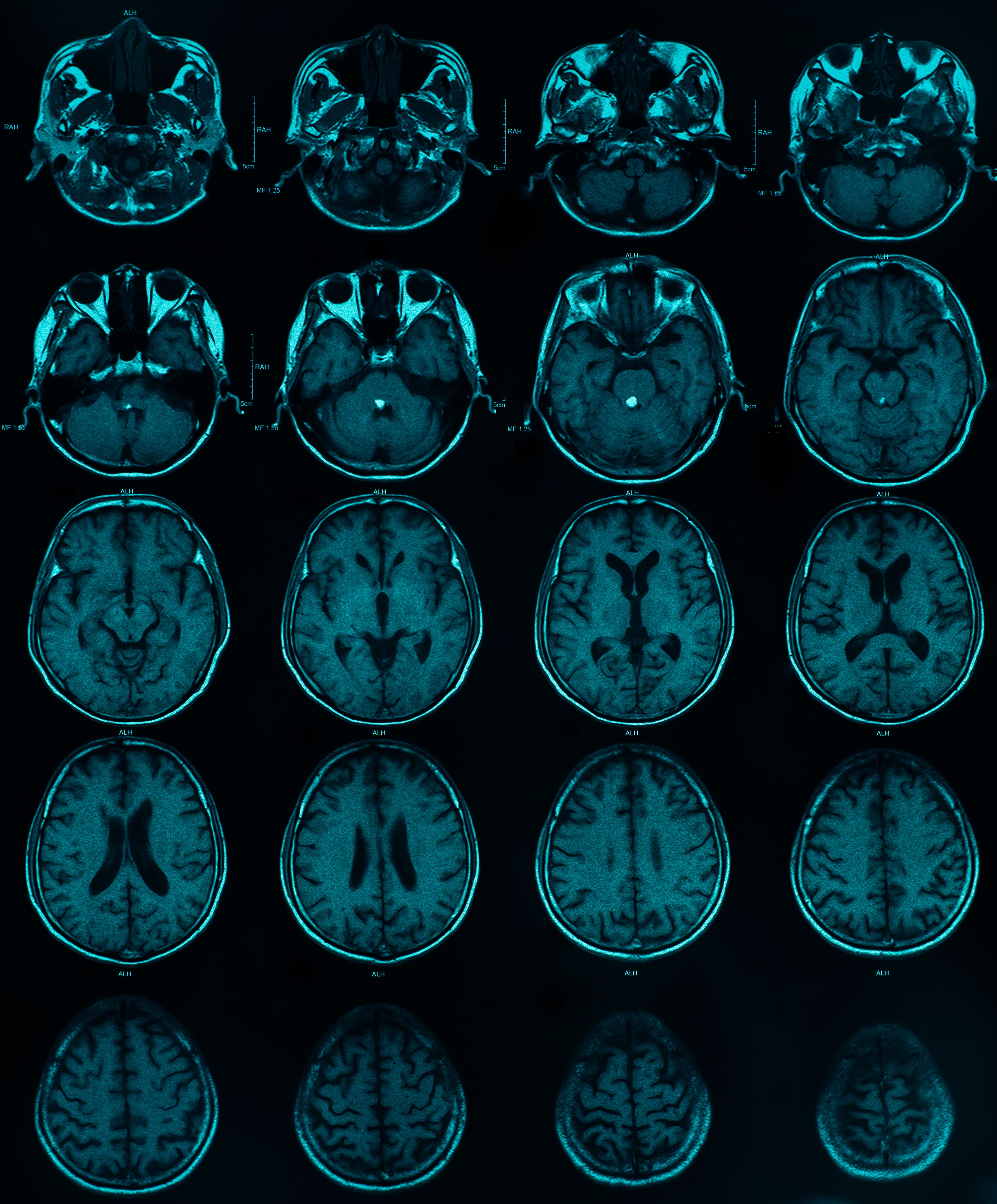
A type of aggressive, treatment-resistant brain tumor has a distinct population of immune cells that support its growth, according to new research led by investigators at the Johns Hopkins Kimmel Cancer Center Bloomberg~Kimmel Institute for Cancer Immunotherapy and the Johns Hopkins University School of Medicine.
Searching for subtypes of immune cells seen only in the most serious, grade 4 brain tumors, called glioblastomas, and using a recently developed technology called spatial genomics, the researchers found that glioblastoma stem cells were co-localized with a type of immunosuppressive cell called myeloid-derived suppressor cell (MDSC), and that these two cells symbiotically feed off of each other to promote tumor growth and aggressiveness. A description of the work was published Jan. 17 in the journal Science.
“Tumor stem cells represent only 5% to 10% of the tumor, but they’re the critical cells that are renewing and generating the rest of the tumor and are essentially responsible for the aggressiveness of the tumor,” says senior study author Drew Pardoll, M.D., Ph.D., the Martin D. Abeloff Professor of Cancer Research , co-director of the Mark Foundation Center for Advanced Genomics and Imaging, and director of the Bloomberg~Kimmel Institute for Cancer Immunotherapy. “We found that the myeloid-derived suppressor cells and tumor stem cells literally were in the same place — a region described by pathologists in the 1980s as the pseudopalisading region. There was a very intimate connection.”
To better characterize the cellular components of brain cancer, investigators performed single-cell RNA sequencing on tissue samples from 33 types of brain tumors spanning from low to high grade, finding two populations of MDSCs in IDH-WT glioblastoma. Then, using a technique called spatial transcriptomics to look at patterns of gene expression of over 750,000 immune cells and more than 350,000 tumor and associated cells in these samples, they found MDSCs were co-located with the tumor stem cells.
“Glioblastoma is a highly aggressive brain tumor with remarkable ability to evade the immune system, which has made immune-based therapies largely ineffective to this point,” said first and co-corresponding author, Christina Jackson, M.D., an assistant professor of neurosurgery at the Perelman School of Medicine at the University of Pennsylvania, who was at Johns Hopkins at the time the research was conducted. “Our study revealed a distinct subset of immune cells, known as myeloid-derived suppressor cells that promote glioblastoma growth, providing new insights into how the tumor interacts with the immune system. By identifying these cells and their role, we hope to uncover new therapeutic targets and lay the groundwork for more effective treatments.”
In their studies, investigators discovered that the two types of cells were feeding each other in the brain tumors. Tumor stem cells were producing chemical signals called chemokines that attracted the MDSCs, and making growth factors and activation factors for the MDSCs. In turn, the MDSCs were producing growth factors for the tumor cells.
Cross-talk between E-MDSCs and tumor cells in IDH-WT glioblastoma

Single-cell RNA sequencing of IDH-WT glioblastoma supports a mechanism of tumor growth involving cross-talk between highly metabolic E-MDSCs and a stem-like subset of glioma cells. In this model, tumor cells produce specific chemokines that attract E-MDSCs to the pseudopalisading region, where rapid growth and oxygen consumption require metabolic adaptation to a high-stress microenvironment. E-MDSCs in turn produce specific growth factors that drive tumor cell growth and aggressiveness.
The researchers were able to further ascertain what specific molecules tumor stem cells were producing to attract and activate MDSCs. Two of the key ones identified by the team were IL (interleukin)-6 and IL-8, which play a role in inflammatory responses, and for which MDSCs have receptors.
“IL-8 is one of the major attractants to bring the MDSCs to the tumor, and IL-6 is one of the major activators of the MDSCs,” Pardoll says.
On the flip side, the team found that MDSCs secreted a growth factor called fibroblast growth factor 11 (FGF11) to feed the stem cells, a molecule never before known to be involved in brain or other cancers.
Along the way, Jackson, Pardoll and colleagues found that tumors with a mutation in the IDH1 gene, which are less aggressive, had almost no MDSCs and far fewer cancer stem cells. This led them to look across all brain cancers at the correlation between MDSC infiltration and survival. Using the National Cancer Institute’s Cancer Genome Atlas (TCGA) database of cancer samples, they indeed found that very tight correlation — the fewer cancer stem cells and fewer MDSCs a person had in their tumors, the better they did.
While additional studies are needed to further understand these cellular interactions, the work is exciting in that it suggests additional potential targets to block in treatment of these aggressive brain tumors, Pardoll says. For example, Jamie Spangler, Ph.D., an associate professor of biomedical engineering at Johns Hopkins, has developed an investigational bispecific antibody that binds to the receptors for IL-6 and IL-8, blocking their signaling.


A type of aggressive, treatment-resistant brain tumor has a distinct population of immune cells that support its growth, according to new research led by investigators at the Johns Hopkins Kimmel Cancer Center Bloomberg~Kimmel Institute for Cancer Immunotherapy and the Johns Hopkins University School of Medicine.
Searching for subtypes of immune cells seen only in the most serious, grade 4 brain tumors, called glioblastomas, and using a recently developed technology called spatial genomics, the researchers found that glioblastoma stem cells were co-localized with a type of immunosuppressive cell called myeloid-derived suppressor cell (MDSC), and that these two cells symbiotically feed off of each other to promote tumor growth and aggressiveness. A description of the work was published Jan. 17 in the journal Science.
To better characterize the cellular components of brain cancer, investigators performed single-cell RNA sequencing on tissue samples from 33 types of brain tumors spanning from low to high grade, finding two populations of MDSCs in IDH-WT glioblastoma. Then, using a technique called spatial transcriptomics to look at patterns of gene expression of over 750,000 immune cells and more than 350,000 tumor and associated cells in these samples, they found MDSCs were co-located with the tumor stem cells.
In their studies, investigators discovered that the two types of cells were feeding each other in the brain tumors. Tumor stem cells were producing chemical signals called chemokines that attracted the MDSCs, and making growth factors and activation factors for the MDSCs. In turn, the MDSCs were producing growth factors for the tumor cells.
Cross-talk between E-MDSCs and tumor cells in IDH-WT glioblastoma
Single-cell RNA sequencing of IDH-WT glioblastoma supports a mechanism of tumor growth involving cross-talk between highly metabolic E-MDSCs and a stem-like subset of glioma cells. In this model, tumor cells produce specific chemokines that attract E-MDSCs to the pseudopalisading region, where rapid growth and oxygen consumption require metabolic adaptation to a high-stress microenvironment. E-MDSCs in turn produce specific growth factors that drive tumor cell growth and aggressiveness.
The researchers were able to further ascertain what specific molecules tumor stem cells were producing to attract and activate MDSCs. Two of the key ones identified by the team were IL (interleukin)-6 and IL-8, which play a role in inflammatory responses, and for which MDSCs have receptors.
On the flip side, the team found that MDSCs secreted a growth factor called fibroblast growth factor 11 (FGF11) to feed the stem cells, a molecule never before known to be involved in brain or other cancers.
Along the way, Jackson, Pardoll and colleagues found that tumors with a mutation in the IDH1 gene, which are less aggressive, had almost no MDSCs and far fewer cancer stem cells. This led them to look across all brain cancers at the correlation between MDSC infiltration and survival. Using the National Cancer Institute’s Cancer Genome Atlas (TCGA) database of cancer samples, they indeed found that very tight correlation — the fewer cancer stem cells and fewer MDSCs a person had in their tumors, the better they did.
While additional studies are needed to further understand these cellular interactions, the work is exciting in that it suggests additional potential targets to block in treatment of these aggressive brain tumors, Pardoll says. For example, Jamie Spangler, Ph.D., an associate professor of biomedical engineering at Johns Hopkins, has developed an investigational bispecific antibody that binds to the receptors for IL-6 and IL-8, blocking their signaling.
Jackson C, Cherry C, Bom S, Dykema AG, Wang R, Thompson E, Zhang M, Li R, Ji Z, Hou W, Zhan W, Zhang H, Choi J, Vaghasia A, Hansen L, Wang W, Bergsneider B, Jones KM, Rodriguez F, Weingart J, Lucas CH, Powell J, Elisseeff J, Yegnasubramanian S, Lim M, Bettegowda C, Ji H, Pardoll D. (2025) Distinct myeloid-derived suppressor cell populations in human glioblastoma. Science 387(6731):eabm5214. [article]
Related Posts
Scientists identify the neuroblastoma cells that signal poor outcomes
Novel biosensor uses DNA ‘barcodes’ to uncover disease signals in cells
CellART extracts single-cell information from high-resolution spatial transcriptomics
Cell2space restores spatial context to single-cell RNA sequencing data
Urine test may improve bladder cancer diagnosis and guide treatment decisions
DeCoRE – a computational method to resolve RNA structural heterogeneity from RNA structure probing data by direct RNA sequencing
Tracing 700 million years of cell evolution
How should biological replicates be handled in long-read RNA sequencing?
RNA-Seq reveals a new way the developing heart controls its growth
The Human RNome Project Consortium seeks to publish first-ever complete sequence of the body’s RNA
scFlowReport brings multiple single-cell RNA sequencing analyses into one workflow
Long-read RNA sequencing reveals how neurons choose alternative RNA endings
DeOPUS improves cell-type estimates from bulk RNA sequencing
ResolVI improves accuracy in spatial transcriptomics analysis
RNA sequencing reveals transcriptional allele differences in prostate cancer stem cells
Unmasking “zombie cells” in aging tissue with an AI-powered barcode
RNA variant imaging for cancer diagnosis
Autism-risk mutations reveal two opposing patterns of brain gene activity
A real-data approach to evaluating RNA transcript analysis methods
Detection and sequencing of Ap2N-capped RNAs in human cells
Stay Connected
Submit a Post to the Blog
Recent Posts
Subscribe to the RNA-Seq Blog
RNA-Seq Products & Services