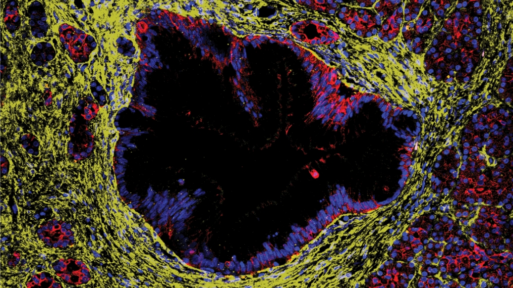
Immunofluorescence image of human donor pancreas with a precursor lesion lined by epithelial cells surrounded by fibroblasts. Credit: Pasca Lab
In an unexpected finding, a new study flips on its head researchers’ understanding of how precancerous pancreas lesions evolve into pancreatic cancer. The paradigm-changing discovery has tremendous implications for identifying people at higher risk of cancer or even, potentially stopping malignant transformation. In tumors, cancer cells induce surrounding non-malignant cells to become “helpers” and promote tumor growth. This is called the microenvironment.
Precursor lesions called pancreatic intraepithelial neoplasia, or PanIN, similarly are surrounded by a collective of other cells. The precursor lesions express a similar set of genes as cancer cells but less strongly. So when researchers looked at the cells in the environment surrounding the lesions, they expected to see the same “tumor light” features. Surprisingly, the microenvironment of the precursor lesions was entirely different.
“It turns out, the microenvironment of these precursor lesions is the same as the microenvironment of the normal pancreas. The lesions have not convinced any of the cells around them to change. That’s not what we were expecting. We were expecting the two components, the cells and the microenvironment, to evolve in lockstep. They did not,” said co-senior study author Marina Pasca di Magliano, Ph.D., Maud T. Lane Professor of Surgical Immunology and Co-director of the Rogel and Blondy Center for Pancreatic Cancer at the University of Michigan.
The study is published in Cancer Discovery, a journal of the American Association for Cancer Research.
This discovery likely explains earlier research from the team that showed precursor lesions are common, including in younger people, while pancreatic cancer remains relatively rare. The transformation of normal pancreas cells into pancreatic cancer has been challenging to study in humans. Microscopic precursor lesions are extremely difficult to isolate in the pancreas and have typically been recovered during surgery when a nearby tumor is removed.
At this point, the PanIN microenvironment is likely influenced by the nearby tumor. The team benefited from a unique collaboration between the U-M Rogel Cancer Center and Gift of Life Michigan, which allows U-M researchers to procure healthy donor pancreases for research. From this partnership, researchers have isolated PanIN lesions from more than 150 donated pancreases from individuals ages 20-70.
For this study, they used multiple cutting edge research technologies, including single cell RNA sequencing and spatial transcriptomics, to isolate single cells, examine them in two dimensions and map the gene expression of specific tissue sections.
“These lesions are like needles in a haystack. The prior way of looking at this was to look at the entire haystack. You get a lot of information about hay and very little information about the needle. These new techniques allow us to just focus in on the needle so we can look at multiple needles using the same amount of computing power and resources,” said co-senior author Timothy Frankel, M.D., Maud T. Lane Professor of Surgical Oncology and co-director of the Rogel and Blondy Center for Pancreatic Cancer at U-M.
Pancreatic cancer has an extensive tumor microenvironment, which includes fibroblasts and immune cells that play complex roles in the biology of the tumor. If precursor lesions are not surrounded by that same microenvironment, it suggests that something else needs to happen to trigger cancer growth – inflammation, pancreatitis, smoking, age, obesity or other stressors linked to pancreatic cancer.
Future studies will seek to understand what factors are involved. The hope is that if researchers can understand how those stressors impact the microenvironment and allow precancerous lesions to turn into cancer, they can target cells involved in that process, intercept them and potentially stop the conversion.
The study reflects the critical role of multidisciplinary team science in pancreatic cancer research. In this case, the robust team at U-M’s Rogel and Blondy Center for Pancreatic Cancer leveraged its unique relationship with Gift of Life and their expertise in pancreatic cancer and its microenvironment, then partnered with additional experts in bioinformatics and pathology at the University of Maryland School of Medicine and New York University.
“It is incredible to see how we can uncover the fundamental cellular mechanisms of disease etiology by blending new computational methods and cutting-edge spatial transcriptomics technologies. Through careful study design, we can use the spatial information to start delving into the unknown dynamics of pancreatic tumor evolution,” said co-corresponding author Elana J. Fertig, Ph.D., Director of the Institute for Genome Sciences at the University of Maryland School of Medicine and Associate Director of Quantitative Sciences at the University of Maryland Marlene and Stewart Greenebaum Comprehensive Cancer Center.
To foster further discovery, the authors have created an open access website and interactive tool to allow other researchers to query the data.
Source – Michigan Medicine


Immunofluorescence image of human donor pancreas with a precursor lesion lined by epithelial cells surrounded by fibroblasts. Credit: Pasca Lab
In an unexpected finding, a new study flips on its head researchers’ understanding of how precancerous pancreas lesions evolve into pancreatic cancer. The paradigm-changing discovery has tremendous implications for identifying people at higher risk of cancer or even, potentially stopping malignant transformation. In tumors, cancer cells induce surrounding non-malignant cells to become “helpers” and promote tumor growth. This is called the microenvironment.
Precursor lesions called pancreatic intraepithelial neoplasia, or PanIN, similarly are surrounded by a collective of other cells. The precursor lesions express a similar set of genes as cancer cells but less strongly. So when researchers looked at the cells in the environment surrounding the lesions, they expected to see the same “tumor light” features. Surprisingly, the microenvironment of the precursor lesions was entirely different.
The study is published in Cancer Discovery, a journal of the American Association for Cancer Research.
This discovery likely explains earlier research from the team that showed precursor lesions are common, including in younger people, while pancreatic cancer remains relatively rare. The transformation of normal pancreas cells into pancreatic cancer has been challenging to study in humans. Microscopic precursor lesions are extremely difficult to isolate in the pancreas and have typically been recovered during surgery when a nearby tumor is removed.
At this point, the PanIN microenvironment is likely influenced by the nearby tumor. The team benefited from a unique collaboration between the U-M Rogel Cancer Center and Gift of Life Michigan, which allows U-M researchers to procure healthy donor pancreases for research. From this partnership, researchers have isolated PanIN lesions from more than 150 donated pancreases from individuals ages 20-70.
For this study, they used multiple cutting edge research technologies, including single cell RNA sequencing and spatial transcriptomics, to isolate single cells, examine them in two dimensions and map the gene expression of specific tissue sections.
Pancreatic cancer has an extensive tumor microenvironment, which includes fibroblasts and immune cells that play complex roles in the biology of the tumor. If precursor lesions are not surrounded by that same microenvironment, it suggests that something else needs to happen to trigger cancer growth – inflammation, pancreatitis, smoking, age, obesity or other stressors linked to pancreatic cancer.
Future studies will seek to understand what factors are involved. The hope is that if researchers can understand how those stressors impact the microenvironment and allow precancerous lesions to turn into cancer, they can target cells involved in that process, intercept them and potentially stop the conversion.
The study reflects the critical role of multidisciplinary team science in pancreatic cancer research. In this case, the robust team at U-M’s Rogel and Blondy Center for Pancreatic Cancer leveraged its unique relationship with Gift of Life and their expertise in pancreatic cancer and its microenvironment, then partnered with additional experts in bioinformatics and pathology at the University of Maryland School of Medicine and New York University.
To foster further discovery, the authors have created an open access website and interactive tool to allow other researchers to query the data.
Source – Michigan Medicine
Elhossiny AM, Kadiyala P, Okoye JO, Hiraki HL, Procario MC, Giridharan T, Watkoske HR, Tannus Ruckert M, Wang J, Griffith BD, Bray AW, Mills JN, Espinoza CE, Zeller J, Peterson N, Bednar F, Zhang Y, Rao A, Lyssiotis CA, Szczepanski JM, Shi J, Deshpande A, Maitra A, Fertig EJ, Carpenter ES, Frankel TL, Pasca di Magliano M. (2026) Asynchronous evolution of epithelium and stroma differentiates precursor lesions from pancreatic cancer. Cancer Discovery. [Epub ahead of print]. [article]
Related Posts
Scientists identify the neuroblastoma cells that signal poor outcomes
GARAGE improves rare cell detection in single-cell RNA sequencing analysis using AI
Novel biosensor uses DNA ‘barcodes’ to uncover disease signals in cells
CellART extracts single-cell information from high-resolution spatial transcriptomics
Cell2space restores spatial context to single-cell RNA sequencing data
Urine test may improve bladder cancer diagnosis and guide treatment decisions
DeCoRE – a computational method to resolve RNA structural heterogeneity from RNA structure probing data by direct RNA sequencing
Tracing 700 million years of cell evolution
How should biological replicates be handled in long-read RNA sequencing?
RNA-Seq reveals a new way the developing heart controls its growth
The Human RNome Project Consortium seeks to publish first-ever complete sequence of the body’s RNA
scFlowReport brings multiple single-cell RNA sequencing analyses into one workflow
Long-read RNA sequencing reveals how neurons choose alternative RNA endings
DeOPUS improves cell-type estimates from bulk RNA sequencing
ResolVI improves accuracy in spatial transcriptomics analysis
RNA sequencing reveals transcriptional allele differences in prostate cancer stem cells
Unmasking “zombie cells” in aging tissue with an AI-powered barcode
RNA variant imaging for cancer diagnosis
Autism-risk mutations reveal two opposing patterns of brain gene activity
A real-data approach to evaluating RNA transcript analysis methods
Stay Connected
Submit a Post to the Blog
Recent Posts
Subscribe to the RNA-Seq Blog
RNA-Seq Products & Services