Scientists analyzed individual cells isolated from two craniopharyngioma subtypes to identify the specific cell types, their features, and their crosstalk
Craniopharyngiomas are brain tumors that negatively impact the hormonal function of the nearby pituitary. The tumor location often prevents necessary surgical intervention. Alternative pharmacological therapy requires an in-depth understanding of the tumor molecular characteristics. To address this gap, researchers from Chiba University analyzed gene expression within individual tumor cells. This study reports the molecular features and interactions of tumor and immune cells associated with two craniopharyngioma subtypes that will help identify future targeted therapeutics.
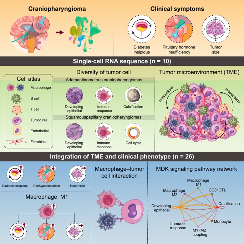
Craniopharyngioma (CP) is a rare brain tumor that develops in the regions close to the hypothalamus and pituitary gland. The CP tumors lead to complications like defective vision, neuronal defects, diabetes, and developmental problems. There are two primary subtypes of CPs: adamantinomatous craniopharyngioma (ACP) and papillary craniopharyngioma (PCP). These two subtypes are distinguished by their distinct genetic profiles. ACP is typically characterized by mutations in the CTNNB1 gene, while PCP is primarily associated with BRAF gene mutations.
The primary course of action for treating CP is surgical intervention. However, the tumor’s invasive nature and its location near critical structures present a significant challenge in terms of surgical intervention. As the tumor progresses, it infiltrates the surrounding tissue, resulting in significant neurological impairments. Therefore, surgery alone is insufficient to address the complex challenges posed by CPs. It is essential to have a comprehensive understanding of the tumor’s biological characteristics and molecular progression in order to ensure successful tumor extraction while preserving surrounding healthy tissue.
Against this backdrop, Professor Tomoaki Tanaka collaborated with Professor Yoshinori Higuchi and Dr. Takashi Kono from the Graduate School of Medicine at Chiba University in Japan to conduct a study to elucidate the underlying biological processes involved in this tumor. The study was made available online on September 30, 2024, and was published in Volume 27 Issue 11 of the journal iScience on November 15, 2024.
To this end, they utilized single-cell RNA sequencing, a technique that reveals differences in gene expression across individual cells, and analyzed 10 cases of CPs.
In an interview, Prof. Tanaka, the senior author of the study, explained the motivation behind it. He said, “Despite being histologically benign, these tumors can significantly impact critical brain structures.” “Our goal was to develop more targeted and less invasive therapeutic approaches that could significantly improve patient outcomes and quality of life.”
The single-cell analysis revealed a diverse range of cell types within the tumor microenvironment (TME), including tumor cells, immune cells, and fibroblasts, with varying proportions across cases. The tumor cells were classified into two main subtypes: Type 1, which is predominant in ACP, and Type 2, which is dominant in PCP. The single-cell gene expression data from the ACP and PCP subtypes was clustered to reveal distinct cell types within the tumors. The study identified cell types linked to the development of epithelial cells and the immune response in both ACP and PCP tumors. However, the cell types involved in tumor calcification were particularly prevalent in ACP, while the cell cycle-associated genes were predominant in the PCP type.
Further, the research team observed a notable diversity in macrophage types between the two tumor types. The pro-inflammatory M1 macrophages and inflammation-related markers were found to be higher in ACP, while the anti-inflammatory M2 macrophages were higher in PCP. Accordingly, a higher ratio of M1 and M2 macrophages was correlated with the occurrence of diabetes and pituitary insufficiency.
Additionally, the study identified a prominent cell-cell interaction between cell surface proteins CD44-secreted phosphoprotein 1 (SPP1). This SPP1–CD44 signal from classical M2 inhibited the sustained proliferation of T cells.
This study presents a comprehensive map of cell types within CP tumors and reports a significant relationship between immune cells and clinical symptoms.
Prof. Tanaka highlighted the clinical implications of their findings, stating, “These findings open up the possibility of personalized treatment approaches for patients with CP based on their specific tumor subtype and immune cell composition. Understanding these differences will also assist clinicians in predicting which patients are at higher risk for complications like diabetes insipidus.”
Going ahead, these findings can enable the creation of new targeted therapies that precisely influence the tumor microenvironment and immune cell interactions, ultimately leading to more effective treatments with reduced adverse effects.
Source – Chiba University


Scientists analyzed individual cells isolated from two craniopharyngioma subtypes to identify the specific cell types, their features, and their crosstalk
Craniopharyngiomas are brain tumors that negatively impact the hormonal function of the nearby pituitary. The tumor location often prevents necessary surgical intervention. Alternative pharmacological therapy requires an in-depth understanding of the tumor molecular characteristics. To address this gap, researchers from Chiba University analyzed gene expression within individual tumor cells. This study reports the molecular features and interactions of tumor and immune cells associated with two craniopharyngioma subtypes that will help identify future targeted therapeutics.
Craniopharyngioma (CP) is a rare brain tumor that develops in the regions close to the hypothalamus and pituitary gland. The CP tumors lead to complications like defective vision, neuronal defects, diabetes, and developmental problems. There are two primary subtypes of CPs: adamantinomatous craniopharyngioma (ACP) and papillary craniopharyngioma (PCP). These two subtypes are distinguished by their distinct genetic profiles. ACP is typically characterized by mutations in the CTNNB1 gene, while PCP is primarily associated with BRAF gene mutations.
The primary course of action for treating CP is surgical intervention. However, the tumor’s invasive nature and its location near critical structures present a significant challenge in terms of surgical intervention. As the tumor progresses, it infiltrates the surrounding tissue, resulting in significant neurological impairments. Therefore, surgery alone is insufficient to address the complex challenges posed by CPs. It is essential to have a comprehensive understanding of the tumor’s biological characteristics and molecular progression in order to ensure successful tumor extraction while preserving surrounding healthy tissue.
Against this backdrop, Professor Tomoaki Tanaka collaborated with Professor Yoshinori Higuchi and Dr. Takashi Kono from the Graduate School of Medicine at Chiba University in Japan to conduct a study to elucidate the underlying biological processes involved in this tumor. The study was made available online on September 30, 2024, and was published in Volume 27 Issue 11 of the journal iScience on November 15, 2024.
To this end, they utilized single-cell RNA sequencing, a technique that reveals differences in gene expression across individual cells, and analyzed 10 cases of CPs.
The single-cell analysis revealed a diverse range of cell types within the tumor microenvironment (TME), including tumor cells, immune cells, and fibroblasts, with varying proportions across cases. The tumor cells were classified into two main subtypes: Type 1, which is predominant in ACP, and Type 2, which is dominant in PCP. The single-cell gene expression data from the ACP and PCP subtypes was clustered to reveal distinct cell types within the tumors. The study identified cell types linked to the development of epithelial cells and the immune response in both ACP and PCP tumors. However, the cell types involved in tumor calcification were particularly prevalent in ACP, while the cell cycle-associated genes were predominant in the PCP type.
Further, the research team observed a notable diversity in macrophage types between the two tumor types. The pro-inflammatory M1 macrophages and inflammation-related markers were found to be higher in ACP, while the anti-inflammatory M2 macrophages were higher in PCP. Accordingly, a higher ratio of M1 and M2 macrophages was correlated with the occurrence of diabetes and pituitary insufficiency.
Additionally, the study identified a prominent cell-cell interaction between cell surface proteins CD44-secreted phosphoprotein 1 (SPP1). This SPP1–CD44 signal from classical M2 inhibited the sustained proliferation of T cells.
This study presents a comprehensive map of cell types within CP tumors and reports a significant relationship between immune cells and clinical symptoms.
Going ahead, these findings can enable the creation of new targeted therapies that precisely influence the tumor microenvironment and immune cell interactions, ultimately leading to more effective treatments with reduced adverse effects.
Source – Chiba University
Matsuda T, Kono T, Taki Y, Sakuma I, Fujimoto M, Hashimoto N, Kawakami E, Fukuhara N, Nishioka H, Inoshita N, Yamada S, Nakamura Y, Horiguchi K, Miki T, Higuchi Y, Tanaka T. (2024) Deciphering craniopharyngioma subtypes: Single-cell analysis of tumor microenvironment and immune networks. iScience 27(11):111068. [article]
Related Posts
RNAmel simplifies RNA sequencing data analysis with a reproducible, integrated platform
Heart failure reshapes bone marrow to create inflammation-related immune memory
Overlooked DNA regions surrounding ‘microexons’ hold missing pieces of autism’s genetic puzzle
RiboBA improves detection of hidden protein-coding regions using ribosome profiling
An anti-senescence gene may fight spinal disc deterioration
Scientists identify the neuroblastoma cells that signal poor outcomes
Novel biosensor uses DNA ‘barcodes’ to uncover disease signals in cells
CellART extracts single-cell information from high-resolution spatial transcriptomics
Cell2space restores spatial context to single-cell RNA sequencing data
Urine test may improve bladder cancer diagnosis and guide treatment decisions
DeCoRE – a computational method to resolve RNA structural heterogeneity from RNA structure probing data by direct RNA sequencing
Tracing 700 million years of cell evolution
How should biological replicates be handled in long-read RNA sequencing?
RNA-Seq reveals a new way the developing heart controls its growth
The Human RNome Project Consortium seeks to publish first-ever complete sequence of the body’s RNA
scFlowReport brings multiple single-cell RNA sequencing analyses into one workflow
Long-read RNA sequencing reveals how neurons choose alternative RNA endings
DeOPUS improves cell-type estimates from bulk RNA sequencing
ResolVI improves accuracy in spatial transcriptomics analysis
RNA sequencing reveals transcriptional allele differences in prostate cancer stem cells
Stay Connected
Submit a Post to the Blog
Recent Posts
Subscribe to the RNA-Seq Blog
RNA-Seq Products & Services