

Understanding how genes are turned on and off requires more than simply measuring RNA levels. Inside the nucleus, DNA is folded in three dimensions, and this folding helps bring regulatory DNA elements, called cis regulatory elements, into contact with the genes they control. These long-range interactions play a critical role in transcription, the process by which genes are converted into RNA.
Researchers at the Duke University Medical Center have developed a new method called scHiCAR that can measure three layers of gene regulation in the same single cell, RNA expression, open chromatin, and 3D genome organization. This technology was reported by scientists working across multiple institutions, including groups involved in brain and genomics research, and enables simultaneous RNA sequencing, chromatin accessibility profiling, and chromosome conformation capture in individual cells.
scHiCAR uses a combinatorial barcoding strategy to link together information about messenger RNA levels, accessible regulatory DNA regions, and physical interactions between distant parts of the genome. Compared to earlier single-cell 3D genome approaches, scHiCAR more efficiently captures long-range interactions anchored at candidate cis regulatory elements. This makes it especially powerful for identifying enhancer promoter connections that influence gene activity.
scHiCAR simultaneously captures mRNA, chromatin accessibility and 3D genome
with high efficiency and accuracy
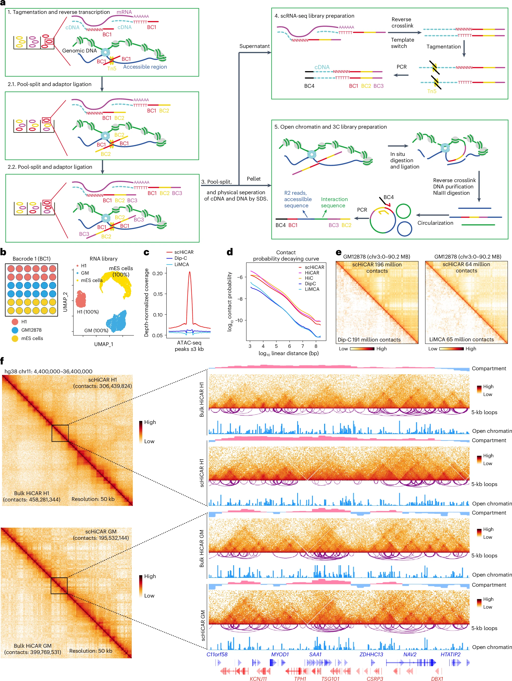
a, Schematics of the scHiCAR experiment workflow. b, Left, BC1 uniquely tags H1 hES cells (red BC1s), human GM12878 cells (blue BC1s) and mES cells (yellow BC1s), serving as the ground truth for cell identity. Right, uniform manifold approximation and projection (UMAP) analysis categorizes cells into three clusters on the basis of their transcriptome data, independent of BC1 information. Subsequent validation with BC1 reveals each cluster’s composition: 100% H1 hES cells, GM12878 cells and mES cells, demonstrating the accuracy of cell identity assignment (accuracy percentages in parentheses). c, The coverage of open chromatin (R2) reads of GM12878 scHiCAR (red), GM12878 Dip-C (blue) and GM12878 LiMCA (light blue) across GM12878 ATAC-seq peaks from ENCODE (ENCFF654BVB). d, Chromatin contact probability (y axis) plotted as a function of linear genomic distance (x axis) measured by scHiCAR (red), HiCAR (purple), in situ Hi-C (orange), Dip-C (blue) and LiMCA (light blue) in GM12878. e, Chromatin contact matrices of scHiCAR with Dip-C (left) and scHiCAR with LiMCA (right) in GM12878. f, Chromatin contact matrices of scHiCAR (above the diagonal) and bulk HiCAR (under the diagonal), with zoomed-in views at chr11:16,600,000–20,640,000 in H1 hES cells (top) and GM12878 (bottom). The right tracks represent the A/B compartment, contact matrices (5 kb bin), Peakachu chromatin interactions (5 kb bin) and open chromatin signals.
The researchers applied scHiCAR to 1.62 million mouse brain cells. With the help of a deep learning based computational tool, they mapped enhancer promoter pairs at 5 kilobase resolution across 22 distinct brain cell types. This allowed them to define cell type specific transcriptomes, accessible regulatory regions, and 3D genome contacts all at once.
The method was also tested in skeletal muscle, a tissue that can be difficult to analyze. In muscle stem cells undergoing regeneration, scHiCAR successfully captured dynamic changes in gene expression, chromatin accessibility, and genome architecture at the single-cell level. This trimodal approach provides a more complete view of how gene regulation changes during tissue repair.
By integrating transcriptome, epigenome, and 3D genome data from the same cells, scHiCAR offers a scalable and cost-effective way to study complex tissues. It reveals how changes in genome folding at specific gene loci contribute to transcriptional control and cell identity.
As single-cell technologies continue to advance, methods like scHiCAR help bridge the gap between DNA structure and gene function, providing deeper insight into development, regeneration, and disease.
Availability – Codes for scHiCAR processing and scDeepLUCIA were deposited to a GitHub repository (https://github.com/DiaoLab/scHiCAR and https://github.com/kaistcbfg/scDeepLUCIA).
Wei X, Xu Y, Yang D, Kim K, Yi L, Luo W, Lin X, Xiang Y, Williams AB, Wang X, Srivas S, Tan C, Zhang K, Li W, Li YE, Yue F, Huang ZJ, Jung I, Diao Y. (2026) Trimodal single-cell profiling of transcriptome, epigenome and 3D genome in complex tissues with scHiCAR. Nature Biotechnology [Epub ahead of print]. [article]
Related Posts






Understanding how genes are turned on and off requires more than simply measuring RNA levels. Inside the nucleus, DNA is folded in three dimensions, and this folding helps bring regulatory DNA elements, called cis regulatory elements, into contact with the genes they control. These long-range interactions play a critical role in transcription, the process by which genes are converted into RNA.
Researchers at the Duke University Medical Center have developed a new method called scHiCAR that can measure three layers of gene regulation in the same single cell, RNA expression, open chromatin, and 3D genome organization. This technology was reported by scientists working across multiple institutions, including groups involved in brain and genomics research, and enables simultaneous RNA sequencing, chromatin accessibility profiling, and chromosome conformation capture in individual cells.
scHiCAR uses a combinatorial barcoding strategy to link together information about messenger RNA levels, accessible regulatory DNA regions, and physical interactions between distant parts of the genome. Compared to earlier single-cell 3D genome approaches, scHiCAR more efficiently captures long-range interactions anchored at candidate cis regulatory elements. This makes it especially powerful for identifying enhancer promoter connections that influence gene activity.
scHiCAR simultaneously captures mRNA, chromatin accessibility and 3D genome
with high efficiency and accuracy
a, Schematics of the scHiCAR experiment workflow. b, Left, BC1 uniquely tags H1 hES cells (red BC1s), human GM12878 cells (blue BC1s) and mES cells (yellow BC1s), serving as the ground truth for cell identity. Right, uniform manifold approximation and projection (UMAP) analysis categorizes cells into three clusters on the basis of their transcriptome data, independent of BC1 information. Subsequent validation with BC1 reveals each cluster’s composition: 100% H1 hES cells, GM12878 cells and mES cells, demonstrating the accuracy of cell identity assignment (accuracy percentages in parentheses). c, The coverage of open chromatin (R2) reads of GM12878 scHiCAR (red), GM12878 Dip-C (blue) and GM12878 LiMCA (light blue) across GM12878 ATAC-seq peaks from ENCODE (ENCFF654BVB). d, Chromatin contact probability (y axis) plotted as a function of linear genomic distance (x axis) measured by scHiCAR (red), HiCAR (purple), in situ Hi-C (orange), Dip-C (blue) and LiMCA (light blue) in GM12878. e, Chromatin contact matrices of scHiCAR with Dip-C (left) and scHiCAR with LiMCA (right) in GM12878. f, Chromatin contact matrices of scHiCAR (above the diagonal) and bulk HiCAR (under the diagonal), with zoomed-in views at chr11:16,600,000–20,640,000 in H1 hES cells (top) and GM12878 (bottom). The right tracks represent the A/B compartment, contact matrices (5 kb bin), Peakachu chromatin interactions (5 kb bin) and open chromatin signals.
The researchers applied scHiCAR to 1.62 million mouse brain cells. With the help of a deep learning based computational tool, they mapped enhancer promoter pairs at 5 kilobase resolution across 22 distinct brain cell types. This allowed them to define cell type specific transcriptomes, accessible regulatory regions, and 3D genome contacts all at once.
The method was also tested in skeletal muscle, a tissue that can be difficult to analyze. In muscle stem cells undergoing regeneration, scHiCAR successfully captured dynamic changes in gene expression, chromatin accessibility, and genome architecture at the single-cell level. This trimodal approach provides a more complete view of how gene regulation changes during tissue repair.
By integrating transcriptome, epigenome, and 3D genome data from the same cells, scHiCAR offers a scalable and cost-effective way to study complex tissues. It reveals how changes in genome folding at specific gene loci contribute to transcriptional control and cell identity.
As single-cell technologies continue to advance, methods like scHiCAR help bridge the gap between DNA structure and gene function, providing deeper insight into development, regeneration, and disease.
Availability – Codes for scHiCAR processing and scDeepLUCIA were deposited to a GitHub repository (https://github.com/DiaoLab/scHiCAR and https://github.com/kaistcbfg/scDeepLUCIA).
Wei X, Xu Y, Yang D, Kim K, Yi L, Luo W, Lin X, Xiang Y, Williams AB, Wang X, Srivas S, Tan C, Zhang K, Li W, Li YE, Yue F, Huang ZJ, Jung I, Diao Y. (2026) Trimodal single-cell profiling of transcriptome, epigenome and 3D genome in complex tissues with scHiCAR. Nature Biotechnology [Epub ahead of print]. [article]
Related Posts
Recent Posts
 Scientists identify the neuroblastoma cells that signal poor outcomes
 GARAGE improves rare cell detection in single-cell RNA sequencing analysis using AI
 Machine learning reveals how RNA splicing is regulated in individual cell types
 Illumina releases SpliceAI2 to help advance rare disease research
 Postdoctoral Fellow – Long-Read RNA Methods Development for Neurodegeneration
 Novel biosensor uses DNA ‘barcodes’ to uncover disease signals in cells
RNA-Seq Products & Services


Stay Connected