Researchers have uncovered what drives the development of scar tissue in the intestines of people with Crohn’s disease.
The study, led by researchers at the Institute of Genetics and Cancer, found clusters of immune cells in the gut which may be stimulating nearby cells to generate excess scar tissue, known as fibrosis.
The discovery could help pinpoint new therapeutic targets to prevent or slow the development of fibrosis, a serious complication of Crohn’s disease.
Crohn’s disease is a chronic inflammatory condition affecting the digestive tract. Over time, ongoing inflammation can lead to fibrosis, where excess collagen builds up in the bowel wall. This scarring can cause the intestine to narrow and become blocked, requiring surgery.
Despite affecting one in five patients with Crohn’s disease, the biological processes that trigger fibrosis remain poorly understood. Current treatments primarily target inflammation rather than the scarring itself.
The research team analysed intestinal tissue samples from Crohn’s disease patients with fibrosis, focusing on the ileum – the final part of the small intestine where the disease most commonly develops.
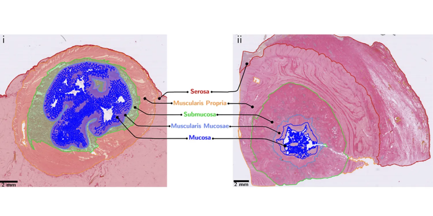
They used archived intestinal tissue samples to examine structural changes across the different layers of the bowel wall.
They found significantly increased fibrosis and immune cell infiltration in Crohn’s disease tissue compared with normal tissue. The submucosa – a deeper layer of the bowel wall – had particularly high levels of scarring, indicating it may play an important role in the early stages of fibrosis.
Researchers next analysed fresh intestinal tissue samples using a cutting-edge technique to study gene activity in individual cells, known as single-cell RNA sequencing.
They identified a link between clusters of immune cells – known as Crohn’s lymphoid aggregates – and groups of endothelial cells, which normally line blood vessels. The endothelial cells appeared to form distinctive structures surrounding the Crohn’s lymphoid aggregates.
Further analysis revealed signalling interactions between these clusters and nearby cells responsible for producing collagen, suggesting that they may actively promote fibrosis.
Our findings highlight previously unrecognised interactions between immune cells, endothelial cells and collagen-producing cells in Crohn’s disease. By combining traditional pathology with single-cell transcriptomics, we were able to confirm these changes using two independent approaches and uncover biological signalling pathways that may provide new therapeutic targets. – Dr Michael GlinkaPostdoctoral Research Fellow
Pathologists, gastroenterologists, biomedical scientists and computer experts at the University of Edinburgh and Heriot-Watt University in Edinburgh worked together with researchers in genomics and transcriptomics at the Earlham Institute and Sanger Institute for over six years, to study the cellular and molecular mechanisms of the process of fibrosis in Crohn’s Disease, in order to identify new therapeutic targets that can be used to treat patients to try to slow down or reverse fibrosis. – Professor Mark ArendsProfessor of Pathology and Head of Edinburgh Pathology, University of Edinburgh
The research highlights potential therapeutic targets that could be explored to interrupt the scarring process and develop treatments specifically aimed at fibrosis in Crohn’s disease.
Fibrosis remains one of the most challenging complications of Crohn’s disease because current treatments primarily target inflammation rather than the scarring itself. Understanding the cellular signalling pathways that link immune activity to collagen production could help guide the development of therapies aimed at preventing or slowing fibrosis. – Dr Shahida DinConsultant Gastroenterologist and Research Clinician at NHS Lothian
Source – The University of Edinburgh


Researchers have uncovered what drives the development of scar tissue in the intestines of people with Crohn’s disease.
The study, led by researchers at the Institute of Genetics and Cancer, found clusters of immune cells in the gut which may be stimulating nearby cells to generate excess scar tissue, known as fibrosis.
The discovery could help pinpoint new therapeutic targets to prevent or slow the development of fibrosis, a serious complication of Crohn’s disease.
Crohn’s disease is a chronic inflammatory condition affecting the digestive tract. Over time, ongoing inflammation can lead to fibrosis, where excess collagen builds up in the bowel wall. This scarring can cause the intestine to narrow and become blocked, requiring surgery.
Despite affecting one in five patients with Crohn’s disease, the biological processes that trigger fibrosis remain poorly understood. Current treatments primarily target inflammation rather than the scarring itself.
The research team analysed intestinal tissue samples from Crohn’s disease patients with fibrosis, focusing on the ileum – the final part of the small intestine where the disease most commonly develops.
They used archived intestinal tissue samples to examine structural changes across the different layers of the bowel wall.
They found significantly increased fibrosis and immune cell infiltration in Crohn’s disease tissue compared with normal tissue. The submucosa – a deeper layer of the bowel wall – had particularly high levels of scarring, indicating it may play an important role in the early stages of fibrosis.
Researchers next analysed fresh intestinal tissue samples using a cutting-edge technique to study gene activity in individual cells, known as single-cell RNA sequencing.
They identified a link between clusters of immune cells – known as Crohn’s lymphoid aggregates – and groups of endothelial cells, which normally line blood vessels. The endothelial cells appeared to form distinctive structures surrounding the Crohn’s lymphoid aggregates.
Further analysis revealed signalling interactions between these clusters and nearby cells responsible for producing collagen, suggesting that they may actively promote fibrosis.
The research highlights potential therapeutic targets that could be explored to interrupt the scarring process and develop treatments specifically aimed at fibrosis in Crohn’s disease.
Source – The University of Edinburgh
Glinka M, Wickham GJ, Nadalin F, Kirkwood KJ, Caldwell H, Wicks M, Hill B, Houghton D, Sharghi M, Kefayat A, Haggarty B, Burger A, Baldock RA, Adams DJ, Papatheodorou I, Bankhead P, Din S, Arends MJ. (2026) Crohn’s lymphoid aggregates with endothelial clusters colocalise with submucosal fibrosis in fibrostenosing Crohn’s disease. Journal of Pathology 268(4): 398-412. [article]
Related Posts
Scientists identify the neuroblastoma cells that signal poor outcomes
Novel biosensor uses DNA ‘barcodes’ to uncover disease signals in cells
CellART extracts single-cell information from high-resolution spatial transcriptomics
Cell2space restores spatial context to single-cell RNA sequencing data
Urine test may improve bladder cancer diagnosis and guide treatment decisions
DeCoRE – a computational method to resolve RNA structural heterogeneity from RNA structure probing data by direct RNA sequencing
Tracing 700 million years of cell evolution
How should biological replicates be handled in long-read RNA sequencing?
RNA-Seq reveals a new way the developing heart controls its growth
The Human RNome Project Consortium seeks to publish first-ever complete sequence of the body’s RNA
scFlowReport brings multiple single-cell RNA sequencing analyses into one workflow
Long-read RNA sequencing reveals how neurons choose alternative RNA endings
DeOPUS improves cell-type estimates from bulk RNA sequencing
ResolVI improves accuracy in spatial transcriptomics analysis
RNA sequencing reveals transcriptional allele differences in prostate cancer stem cells
Unmasking “zombie cells” in aging tissue with an AI-powered barcode
RNA variant imaging for cancer diagnosis
Autism-risk mutations reveal two opposing patterns of brain gene activity
A real-data approach to evaluating RNA transcript analysis methods
Detection and sequencing of Ap2N-capped RNAs in human cells
Stay Connected
Submit a Post to the Blog
Recent Posts
Subscribe to the RNA-Seq Blog
RNA-Seq Products & Services