

Understanding how cells function within tissues requires more than just studying individual molecular layers like gene expression or DNA modifications. Scientists need a way to integrate multiple layers of biological data while preserving the spatial context of cells within their environment. However, current technologies in spatial and single-cell omics sequencing have technical limitations that make this challenging.
To address this, researchers at Zhejiang University have developed SIMO—a computational tool designed to integrate various types of molecular data from different single-cell and spatial sequencing techniques. Unlike previous methods that mainly connect spatial transcriptomics (which maps where genes are active in tissues) with single-cell RNA sequencing, SIMO goes a step further. It can combine multiple data types, including chromatin accessibility (how DNA is packed and exposed for gene activation) and DNA methylation (chemical modifications that influence gene expression), even when they haven’t been spatially measured together before.
Spatial integration of multi-omics single-cell data with SIMO
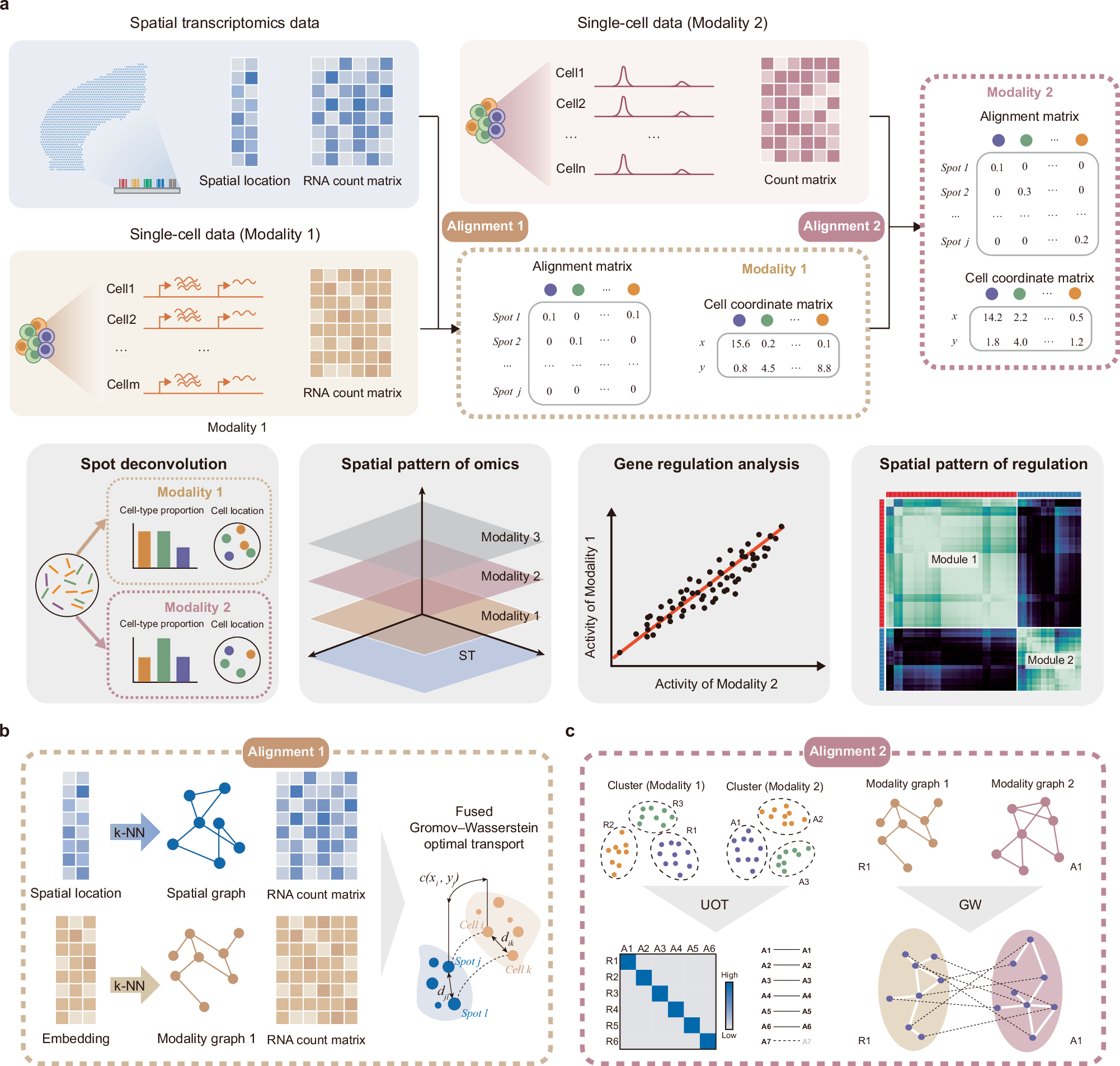
a SIMO takes ST and multi-omics single-cell data as input. SIMO assigns individual cells from various modalities to specific spots, refining the spatial coordinates of single cells grounded on either the similarity of gene expression or the congruence of low-dimensional embedding representations. This process yields a comprehensive spatial multimodal dataset. By assigning coordinates to cells from different modality data, SIMO enables multidimensional deconvolution of spots and reconstruction of spatial omics patterns. Moreover, the downstream functions of SIMO can realize gene regulation analysis and spatial regulation analysis. b By leveraging the fused Gromov-Wasserstein optimal transport algorithm and taking into consideration the gene expression, as well as spatial and modal graphs constructed through k-NN, SIMO computes a probabilistic alignment between cells and spots. c For a second modality, SIMO integrates it with the already mapped transcriptomic data, utilizing Unbalanced Optimal Transport (UOT) to facilitate cluster-based label transfer. Following this, SIMO calculates cell alignment using Gromov-Wasserstein (GW) based on k-NN graphs.
How SIMO Works
SIMO operates through probabilistic alignment, meaning it mathematically matches different types of single-cell and spatial data to create a more complete picture of how cells function within tissues. The researchers tested SIMO on simulated datasets and found that it performed with high accuracy and reliability.
Applying SIMO to real biological data allowed the researchers to uncover topological patterns—how cells are organized in space—and understand their regulatory behavior across multiple molecular layers. This means that SIMO can detect previously hidden spatial heterogeneity, revealing how different biological molecules work together in specific regions of tissues.
Advancing Spatial Biology
By providing a more comprehensive way to analyze spatial omics data, SIMO represents a major advancement in spatial biology. It allows scientists to explore molecular interactions in greater detail, offering new insights into tissue organization, disease progression, and potential therapeutic targets. With SIMO, researchers can uncover multimodal spatial relationships that were previously inaccessible, paving the way for new discoveries in biomedical research.
Availability – The SIMO toolkit is available at GitHub: https://github.com/ZJUFanLab/SIMO
Yang P, Jin K, Yao Y, Jin L, Shao X, Li C, Lu X, Fan X. (2025) Spatial integration of multi-omics single-cell data with SIMO. Nat Commun 16(1):1265. [article]
Related Posts




Understanding how cells function within tissues requires more than just studying individual molecular layers like gene expression or DNA modifications. Scientists need a way to integrate multiple layers of biological data while preserving the spatial context of cells within their environment. However, current technologies in spatial and single-cell omics sequencing have technical limitations that make this challenging.
To address this, researchers at Zhejiang University have developed SIMO—a computational tool designed to integrate various types of molecular data from different single-cell and spatial sequencing techniques. Unlike previous methods that mainly connect spatial transcriptomics (which maps where genes are active in tissues) with single-cell RNA sequencing, SIMO goes a step further. It can combine multiple data types, including chromatin accessibility (how DNA is packed and exposed for gene activation) and DNA methylation (chemical modifications that influence gene expression), even when they haven’t been spatially measured together before.
Spatial integration of multi-omics single-cell data with SIMO
a SIMO takes ST and multi-omics single-cell data as input. SIMO assigns individual cells from various modalities to specific spots, refining the spatial coordinates of single cells grounded on either the similarity of gene expression or the congruence of low-dimensional embedding representations. This process yields a comprehensive spatial multimodal dataset. By assigning coordinates to cells from different modality data, SIMO enables multidimensional deconvolution of spots and reconstruction of spatial omics patterns. Moreover, the downstream functions of SIMO can realize gene regulation analysis and spatial regulation analysis. b By leveraging the fused Gromov-Wasserstein optimal transport algorithm and taking into consideration the gene expression, as well as spatial and modal graphs constructed through k-NN, SIMO computes a probabilistic alignment between cells and spots. c For a second modality, SIMO integrates it with the already mapped transcriptomic data, utilizing Unbalanced Optimal Transport (UOT) to facilitate cluster-based label transfer. Following this, SIMO calculates cell alignment using Gromov-Wasserstein (GW) based on k-NN graphs.
How SIMO Works
SIMO operates through probabilistic alignment, meaning it mathematically matches different types of single-cell and spatial data to create a more complete picture of how cells function within tissues. The researchers tested SIMO on simulated datasets and found that it performed with high accuracy and reliability.
Applying SIMO to real biological data allowed the researchers to uncover topological patterns—how cells are organized in space—and understand their regulatory behavior across multiple molecular layers. This means that SIMO can detect previously hidden spatial heterogeneity, revealing how different biological molecules work together in specific regions of tissues.
Advancing Spatial Biology
By providing a more comprehensive way to analyze spatial omics data, SIMO represents a major advancement in spatial biology. It allows scientists to explore molecular interactions in greater detail, offering new insights into tissue organization, disease progression, and potential therapeutic targets. With SIMO, researchers can uncover multimodal spatial relationships that were previously inaccessible, paving the way for new discoveries in biomedical research.
Availability – The SIMO toolkit is available at GitHub: https://github.com/ZJUFanLab/SIMO
Yang P, Jin K, Yao Y, Jin L, Shao X, Li C, Lu X, Fan X. (2025) Spatial integration of multi-omics single-cell data with SIMO. Nat Commun 16(1):1265. [article]
Related Posts
Recent Posts
 RNAmel simplifies RNA sequencing data analysis with a reproducible, integrated platform
 Heart failure reshapes bone marrow to create inflammation-related immune memory
 Overlooked DNA regions surrounding ‘microexons’ hold missing pieces of autism’s genetic puzzle
 RiboBA improves detection of hidden protein-coding regions using ribosome profiling
 An anti-senescence gene may fight spinal disc deterioration
 Bulk RNA-seq Analysis: A Complete Beginner’s Guide
RNA-Seq Products & Services


Stay Connected