Scientists at the Yale School of Medicine (YSM) are using single-cell RNA sequencing to learn how SARS-CoV-2 interacts with a host cell. The two senior authors David van Dijk, PhD, an assistant professor of medicine in the Section of Cardiovascular Medicine and Computer Science, and Craig Wilen, MD, PhD, an assistant professor of Laboratory Medicine and of Immunobiology, utilized single-cell RNA sequencing of infected human bronchial epithelial cells (HBECs) to determine how the virus infects and alters healthy cells.
In the bioRxiv study the authors identified ciliated cells as the major target of SARS-CoV-2 infection. The bronchial epithelium acts as a protective barrier against allergens and pathogens. Cilia removes mucus and other particles from the respiratory tract. Their findings offer insight into how the virus causes disease.
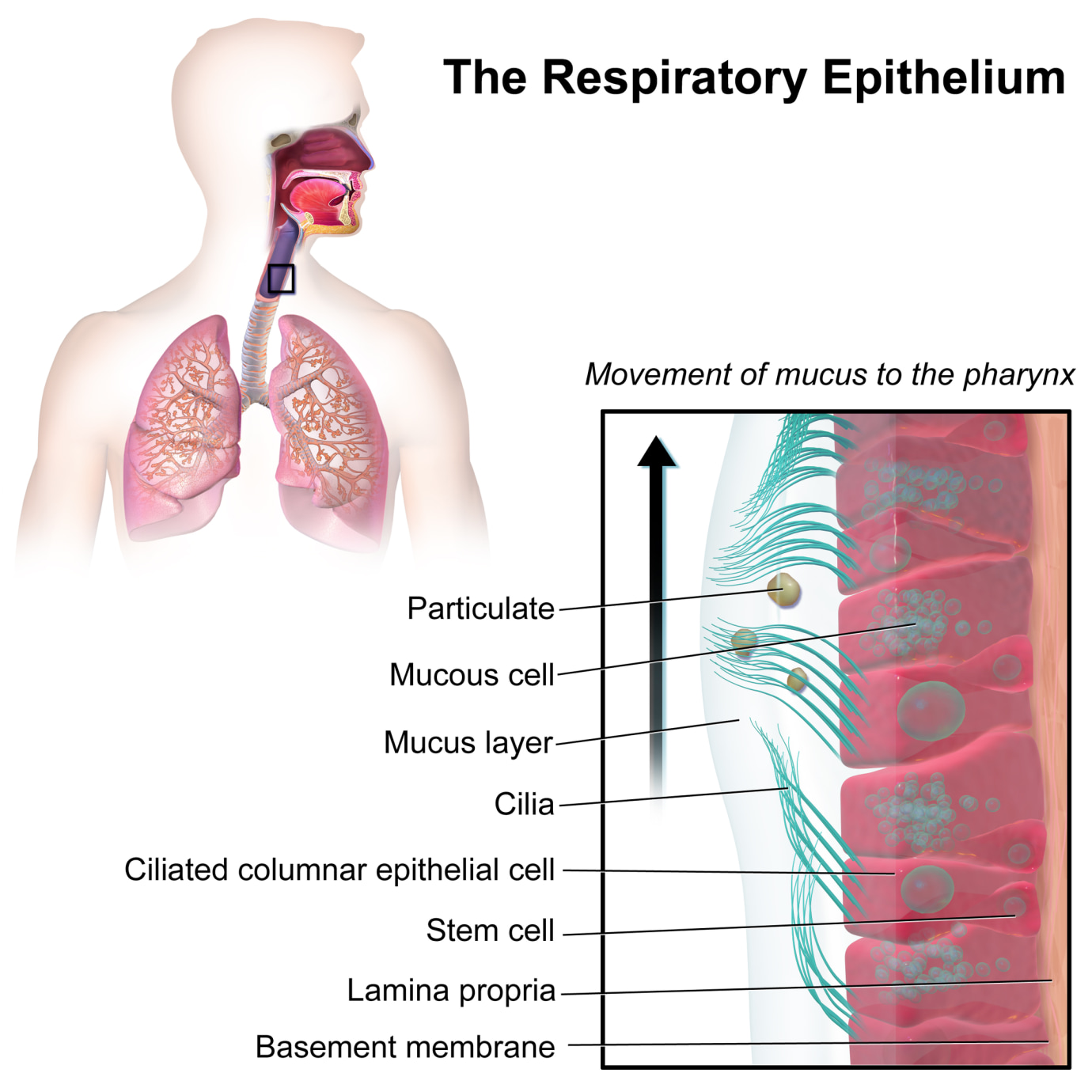
Photo by Blausen Medical
Wilen and post-doctoral associate Mia Alfajaro, PhD, infected HBECs in an air-liquid interface with SARS-CoV-2. Over three days, they used single-cell RNA sequencing to identify signatures of infection dynamics such as the number of infected cells across cell types, and whether SARS-CoV-2 activated an immune response in infected cells. Van Dijk, who specializes in single-cell technologies, utilized advanced algorithms to develop working hypotheses.
“Machine learning allows us to generate hypotheses. It’s a different way of doing science. We go in with as few hypotheses as possible. Measure everything we can measure, and the algorithms present the hypothesis to us,” he said.
Visualization of the experiment
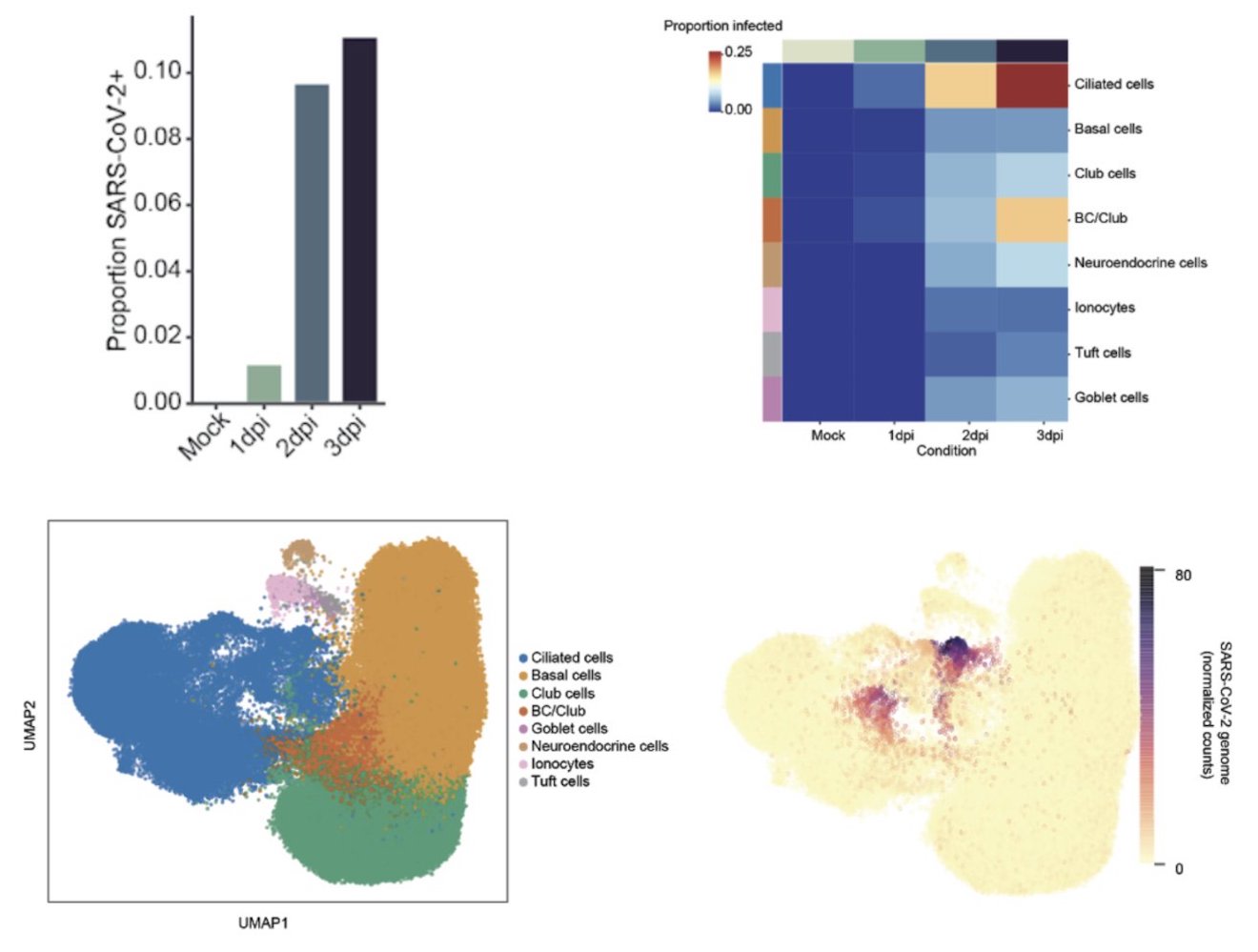
Cultured human bronchial epithelial cells (HBECs) were analyzed 1, 2 and 3 days post infection with SARS-CoV-2. – Photo by David van Dijk and Craig Wilen
The researchers collaborated with Tamas Horvath, PhD, and Klara Szigeti-Buck to use electron microscopy to learn about the structural basis of the virus and target cells. These observations provide insights about host-virus interaction to measure SARS-CoV-2 cell tropism, or the ability of the virus to infect different cell types, as identified by the algorithms. After three days, thousands of cultured cells became infected. The authors analyzed data from the infected cells along with neighboring bystander cells. They observed ciliated cells were 83% of the infected cells. These cells were the first and primary source of infection throughout the study. The virus also targeted other epithelial cell types including basal and club cells. The goblet, neuroendocrine, tuft cells, and ionocytes were less likely to become infected.
The gene signatures revealed an innate immune response associated with a protein called Interleukin 6 (IL-6). The analysis also showed a shift in the polyadenylated viral transcripts. Lastly, the (uninfected) bystander cells also showed an immune response, likely due to signals from the infected cells. Pulling from tens of thousands of genes, the algorithms locate the genetic differences between infected and non-infected cells. They also received a $100,000 grant from fastgrants.org to continue their research.
In the next phase of this study, Wilen and van Dijk will examine the severity of SARS-CoV-2 compared to other types of coronaviruses, and conduct tests in animal models.
Source – Yale School of Medicine
Ravindra NG et al. (2020) Single-cell longitudinal analysis of SARS-CoV-2 infection in human bronchial epithelial cells. bioRXiv [Epub ahead of print]. [abstract]


Scientists at the Yale School of Medicine (YSM) are using single-cell RNA sequencing to learn how SARS-CoV-2 interacts with a host cell. The two senior authors David van Dijk, PhD, an assistant professor of medicine in the Section of Cardiovascular Medicine and Computer Science, and Craig Wilen, MD, PhD, an assistant professor of Laboratory Medicine and of Immunobiology, utilized single-cell RNA sequencing of infected human bronchial epithelial cells (HBECs) to determine how the virus infects and alters healthy cells.
In the bioRxiv study the authors identified ciliated cells as the major target of SARS-CoV-2 infection. The bronchial epithelium acts as a protective barrier against allergens and pathogens. Cilia removes mucus and other particles from the respiratory tract. Their findings offer insight into how the virus causes disease.
Photo by Blausen Medical
Wilen and post-doctoral associate Mia Alfajaro, PhD, infected HBECs in an air-liquid interface with SARS-CoV-2. Over three days, they used single-cell RNA sequencing to identify signatures of infection dynamics such as the number of infected cells across cell types, and whether SARS-CoV-2 activated an immune response in infected cells. Van Dijk, who specializes in single-cell technologies, utilized advanced algorithms to develop working hypotheses.
Visualization of the experiment
Cultured human bronchial epithelial cells (HBECs) were analyzed 1, 2 and 3 days post infection with SARS-CoV-2. – Photo by David van Dijk and Craig Wilen
The researchers collaborated with Tamas Horvath, PhD, and Klara Szigeti-Buck to use electron microscopy to learn about the structural basis of the virus and target cells. These observations provide insights about host-virus interaction to measure SARS-CoV-2 cell tropism, or the ability of the virus to infect different cell types, as identified by the algorithms. After three days, thousands of cultured cells became infected. The authors analyzed data from the infected cells along with neighboring bystander cells. They observed ciliated cells were 83% of the infected cells. These cells were the first and primary source of infection throughout the study. The virus also targeted other epithelial cell types including basal and club cells. The goblet, neuroendocrine, tuft cells, and ionocytes were less likely to become infected.
The gene signatures revealed an innate immune response associated with a protein called Interleukin 6 (IL-6). The analysis also showed a shift in the polyadenylated viral transcripts. Lastly, the (uninfected) bystander cells also showed an immune response, likely due to signals from the infected cells. Pulling from tens of thousands of genes, the algorithms locate the genetic differences between infected and non-infected cells. They also received a $100,000 grant from fastgrants.org to continue their research.
In the next phase of this study, Wilen and van Dijk will examine the severity of SARS-CoV-2 compared to other types of coronaviruses, and conduct tests in animal models.
Source – Yale School of Medicine
Ravindra NG et al. (2020) Single-cell longitudinal analysis of SARS-CoV-2 infection in human bronchial epithelial cells. bioRXiv [Epub ahead of print]. [abstract]
Related Posts
Researchers create first whole-body single-cell atlas of an adult vertebrate
UAlbany Joins $10 Million Effort to Map RNA Modifications in the Human Body
Overlooked ‘off switch’ in lung tumors may explain why immunotherapy doesn’t work for everyone
Squids Edit RNA for Novel Protein Functions
New marker panel and web tool help define early human cell fates
Researchers Develop AI Tool to Map MicroRNA Binding Sites on Circular RNAs
“Raman Genome” for SARS-CoV-2: new Raman technology distinguishes viral variants by their molecular RNA architecture
Advanced paternal age has sex-specific effects on fetal brain gene expression
Exercise is good for us—but why?
Heart failure reshapes bone marrow to create inflammation-related immune memory
Overlooked DNA regions surrounding ‘microexons’ hold missing pieces of autism’s genetic puzzle
An anti-senescence gene may fight spinal disc deterioration
Scientists identify the neuroblastoma cells that signal poor outcomes
Urine test may improve bladder cancer diagnosis and guide treatment decisions
Tracing 700 million years of cell evolution
RNA-Seq reveals a new way the developing heart controls its growth
The Human RNome Project Consortium seeks to publish first-ever complete sequence of the body’s RNA
Unmasking “zombie cells” in aging tissue with an AI-powered barcode
RNA variant imaging for cancer diagnosis
Autism-risk mutations reveal two opposing patterns of brain gene activity
Stay Connected
Submit a Post to the Blog
Recent Posts
Subscribe to the RNA-Seq Blog
RNA-Seq Products & Services