

Glioblastoma (GBM) is one of the most aggressive and difficult-to-treat brain cancers. Scientists have learned that not all GBMs are the same—they can vary significantly based on their tumor microenvironment (TME), which includes the surrounding cells, molecules, and blood vessels that support tumor growth. Understanding these differences is essential for developing treatments that target specific GBM subtypes.
A research team at the University of Salamanca has used single-nucleus RNA sequencing (snRNA-seq) to examine how tumor cells and their microenvironment interact in a commonly used mouse model of GBM. By analyzing tumors at various stages of growth and treatment, the team identified key features that mirror those found in the most common subtype of human GBM—known as TMEMed GBM.
Study overview
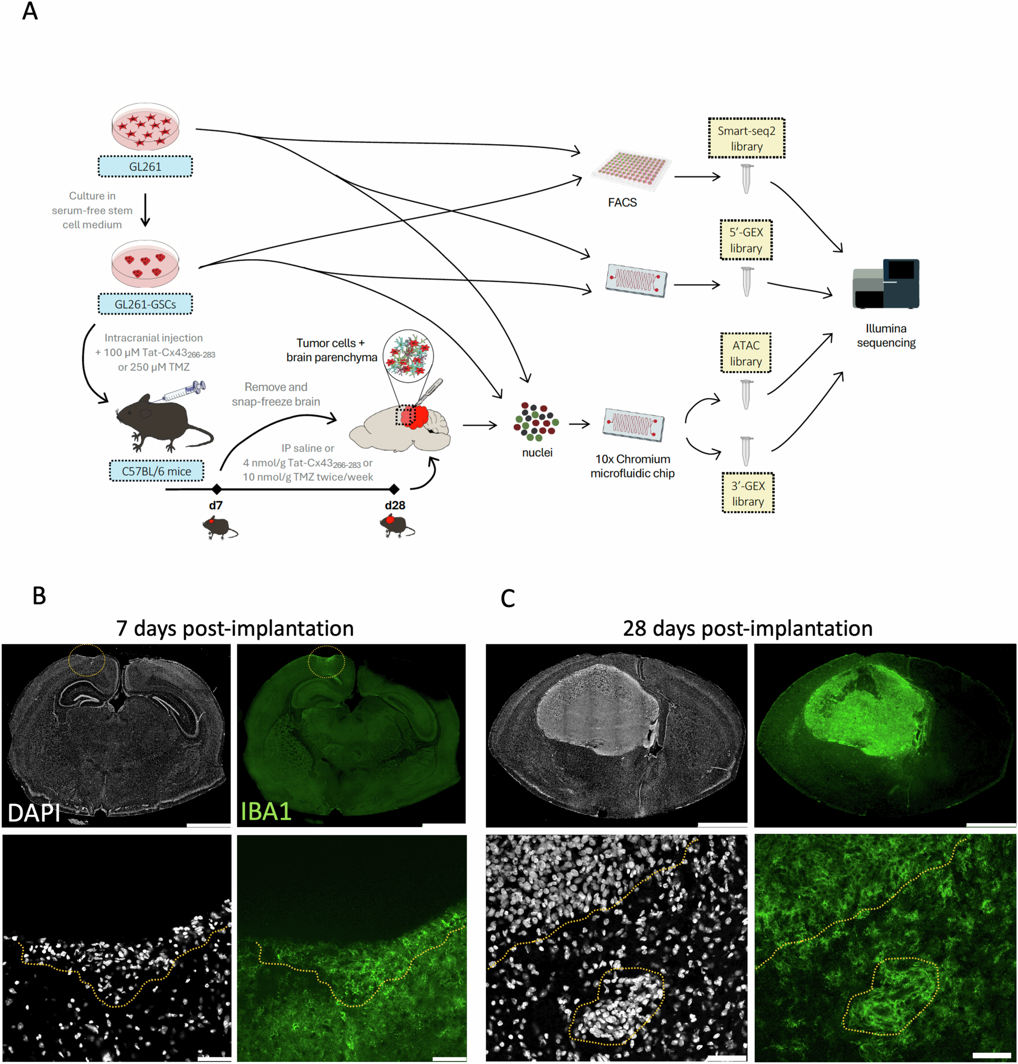
A GL261-GSCs were grown as neurospheres in stem cell medium. They were obtained from GL261 cells grown in adherence in differentiation medium. 5000 GL261-GSCs were intracranially injected into the right hemisphere of brains of syngeneic C57BL/6 mice in saline solution or 100 µM Tat-Cx43266-283 (Tat-Cx43) or 250 µM temozolomide (TMZ). After 7 days, these treatments were intraperitoneally (IP) administered twice per week. Samples were collected from GBM-bearing mouse brains, including tumor core and surrounding tissue, at two stages of tumor growth, early (7 days post-implantation) and late (28 days post-implantation), and isolated nuclei for snRNA-Seq. snRNA-Seq was performed in all the in vitro and in vivo samples. Created with Bioicons: 96_well_plate and and microfluids_chip icons by Marcel Tisch licensed under CC0. B Illustrative IBA1 immunofluorescence images of brains at 7 days post-implantation showing a small group of DAPI-stained tumor cells surrounded by IBA1+ TAMs at the injection site (dashed circle in upper panels), scale bar: 2 mm. Lower panels: zoomed-in images showing tumor cells intermingled and surrounded by IBA1+ TAMs (dashed line), scale bar: 50 µm. C Illustrative IBA1 immunofluorescence images of brains at 28 days post-implantation showing large tumors infiltrated and surrounded by IBA1+ TAMs, scale bar: 2 mm. Lower panels: zoomed-in images showing the border of the tumor together with a group of tumor cells infiltrating the brain parenchyma (dashed line), scale bar: 50 µm. Note the abundance of IBA1+ TAMs infiltrating and surrounding these tumors.
To further validate their findings, the researchers used a technique called spatial transcriptomics, which allowed them to see exactly where different cell types were located in the tumor. This confirmed that the mouse model shares important molecular and cellular characteristics with human TMEMed GBM.
Notably, the model revealed specific genes involved in how tumor cells connect to neural circuits, evade the immune system, and suppress immune responses. Some of these genes—including Grik2, Cd274 (PD-L1), and Mrc1—are also potential targets for immunotherapy, a treatment strategy that helps the immune system fight cancer.
By showing that this mouse model closely mimics the most frequent GBM subtype in humans, these researchers have provided researchers with a valuable tool for testing new therapies with greater precision and relevance to human disease.
García-Vicente L, Martínez-Fernández M, Borja M, Tran V, Álvarez-Vázquez A, Flores-Hernández R, Ding Y, González-Sánchez R, Granados A, McGeever E, Kim YJ, Detweiler A, Mekonen H, Paul S, Pisco AO, Neff NF, Tabernero A. (2025) Single-nucleus RNA sequencing reveals a preclinical model for the most common subtype of glioblastoma. Commun Biol 8(1):671. [article]
Related Posts




Glioblastoma (GBM) is one of the most aggressive and difficult-to-treat brain cancers. Scientists have learned that not all GBMs are the same—they can vary significantly based on their tumor microenvironment (TME), which includes the surrounding cells, molecules, and blood vessels that support tumor growth. Understanding these differences is essential for developing treatments that target specific GBM subtypes.
A research team at the University of Salamanca has used single-nucleus RNA sequencing (snRNA-seq) to examine how tumor cells and their microenvironment interact in a commonly used mouse model of GBM. By analyzing tumors at various stages of growth and treatment, the team identified key features that mirror those found in the most common subtype of human GBM—known as TMEMed GBM.
Study overview
A GL261-GSCs were grown as neurospheres in stem cell medium. They were obtained from GL261 cells grown in adherence in differentiation medium. 5000 GL261-GSCs were intracranially injected into the right hemisphere of brains of syngeneic C57BL/6 mice in saline solution or 100 µM Tat-Cx43266-283 (Tat-Cx43) or 250 µM temozolomide (TMZ). After 7 days, these treatments were intraperitoneally (IP) administered twice per week. Samples were collected from GBM-bearing mouse brains, including tumor core and surrounding tissue, at two stages of tumor growth, early (7 days post-implantation) and late (28 days post-implantation), and isolated nuclei for snRNA-Seq. snRNA-Seq was performed in all the in vitro and in vivo samples. Created with Bioicons: 96_well_plate and and microfluids_chip icons by Marcel Tisch licensed under CC0. B Illustrative IBA1 immunofluorescence images of brains at 7 days post-implantation showing a small group of DAPI-stained tumor cells surrounded by IBA1+ TAMs at the injection site (dashed circle in upper panels), scale bar: 2 mm. Lower panels: zoomed-in images showing tumor cells intermingled and surrounded by IBA1+ TAMs (dashed line), scale bar: 50 µm. C Illustrative IBA1 immunofluorescence images of brains at 28 days post-implantation showing large tumors infiltrated and surrounded by IBA1+ TAMs, scale bar: 2 mm. Lower panels: zoomed-in images showing the border of the tumor together with a group of tumor cells infiltrating the brain parenchyma (dashed line), scale bar: 50 µm. Note the abundance of IBA1+ TAMs infiltrating and surrounding these tumors.
To further validate their findings, the researchers used a technique called spatial transcriptomics, which allowed them to see exactly where different cell types were located in the tumor. This confirmed that the mouse model shares important molecular and cellular characteristics with human TMEMed GBM.
Notably, the model revealed specific genes involved in how tumor cells connect to neural circuits, evade the immune system, and suppress immune responses. Some of these genes—including Grik2, Cd274 (PD-L1), and Mrc1—are also potential targets for immunotherapy, a treatment strategy that helps the immune system fight cancer.
By showing that this mouse model closely mimics the most frequent GBM subtype in humans, these researchers have provided researchers with a valuable tool for testing new therapies with greater precision and relevance to human disease.
García-Vicente L, Martínez-Fernández M, Borja M, Tran V, Álvarez-Vázquez A, Flores-Hernández R, Ding Y, González-Sánchez R, Granados A, McGeever E, Kim YJ, Detweiler A, Mekonen H, Paul S, Pisco AO, Neff NF, Tabernero A. (2025) Single-nucleus RNA sequencing reveals a preclinical model for the most common subtype of glioblastoma. Commun Biol 8(1):671. [article]
Related Posts
Recent Posts
 Scientists identify the neuroblastoma cells that signal poor outcomes
 GARAGE improves rare cell detection in single-cell RNA sequencing analysis using AI
 Machine learning reveals how RNA splicing is regulated in individual cell types
 Illumina releases SpliceAI2 to help advance rare disease research
 Postdoctoral Fellow – Long-Read RNA Methods Development for Neurodegeneration
 Novel biosensor uses DNA ‘barcodes’ to uncover disease signals in cells
RNA-Seq Products & Services



Stay Connected