

Understanding how cells are arranged in tissues is just as important as knowing what genes they are expressing. Traditional single-cell and single-nucleus RNA sequencing has given researchers valuable insights into gene activity, but until now, it has not been easy to pinpoint exactly where those cells are located in tissue.
To solve this problem, researchers at the Broad Institute of Harvard and MIT developed a new method called Slide-tags. This technique attaches special DNA barcodes to individual nuclei in intact tissue slices. These barcodes act like precise “location tags,” allowing scientists to link high-quality RNA sequencing and other molecular data with the exact position of each cell in a tissue.
When applied to the mouse hippocampus, Slide-tags provided spatial resolution of less than 10 micrometers while maintaining full transcriptome quality. The researchers also successfully used it on human tissues such as brain, tonsil, and melanoma, showing how the approach can reveal spatially distinct patterns of gene activity. For example, they were able to uncover how immune cells interact during B cell maturation and how cancer cells change state in different parts of a tumor.
Slide-tags enables single-nucleus spatial transcriptomics in the mouse hippocampus
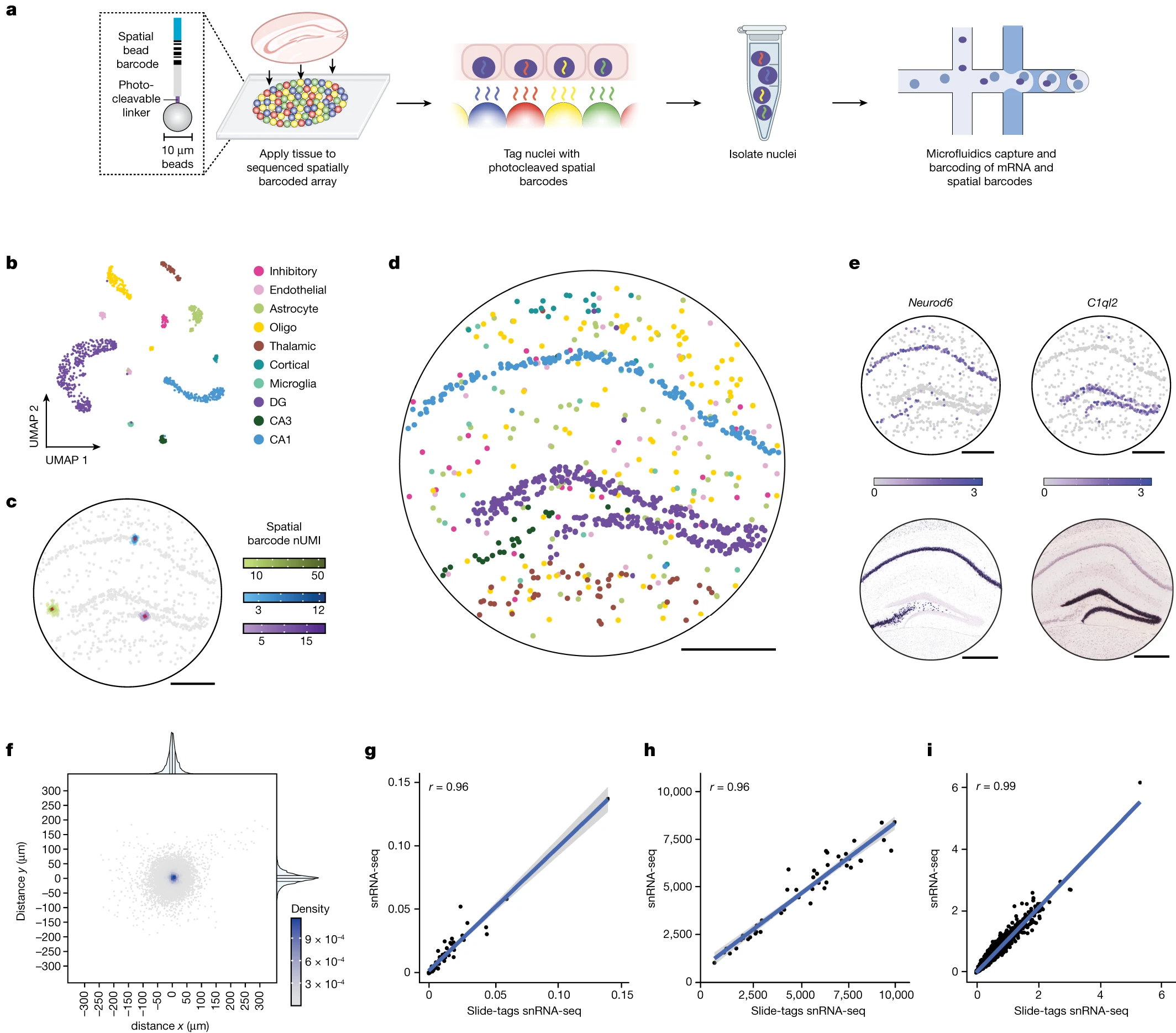
a, Schematic of Slide-tags. b, Uniform manifold approximation and projection (UMAP) embedding of snRNA-seq profiles coloured by cell type annotations. DG, dentate gyrus; oligo, oligodendrocyte. c, The signal spatial barcode clusters after noise filtering by DBSCAN for selected cells, coloured according to cell type annotations (as in b) and the number of spatial barcode UMIs. Raw plots for these cells are plotted in Extended Data Fig. 2k d, Slide-tags enables localization of nuclei to spatial coordinates in the mouse hippocampus; cells are coloured according to cell type annotation (as in b). e, Spatial expression of known marker genes compared with in situ hybridization data from the Allen Mouse Brain Atlas. Colour scales, normalized average counts. f, The distance from the centroid for each of the spatial barcodes across all signal spatial barcode clusters. g–i, Comparison metrics plotted for snRNA-seq compared with Slide-tags snRNA-seq, performed on consecutive sections. g, Cell type proportions and mean UMIs per cell are plotted by cell type. h, The normalized average UMI counts were determined per gene across all cells. i, Normalized average counts.
One of the biggest advantages of Slide-tags is flexibility. It can be integrated with many existing single-cell techniques, including measurements of chromatin accessibility, RNA, and even T cell receptor sequences. This means scientists now have a powerful universal tool for bringing the full scope of single-cell technologies into the world of spatial genomics.
Availability – Code for processing spatial sequencing libraries is available at GitHub (https://github.com/broadchenf/Slide-tags
Russell A, Weir J, Nadaf N, Shabet M, Kumar V, Kambhampati S, Raichur R, Marrero G, Liu S, Balderrama K, Vanderburg C, Shanmugam V, Tian L, Iorgulescu J, Yoon C, Wu C, Macosko E, Chen F. (2024) Slide-tags enables single-nucleus barcoding for multimodal spatial genomics. Nature 625(7993): 101-109. [article]
Related Posts




Understanding how cells are arranged in tissues is just as important as knowing what genes they are expressing. Traditional single-cell and single-nucleus RNA sequencing has given researchers valuable insights into gene activity, but until now, it has not been easy to pinpoint exactly where those cells are located in tissue.
To solve this problem, researchers at the Broad Institute of Harvard and MIT developed a new method called Slide-tags. This technique attaches special DNA barcodes to individual nuclei in intact tissue slices. These barcodes act like precise “location tags,” allowing scientists to link high-quality RNA sequencing and other molecular data with the exact position of each cell in a tissue.
When applied to the mouse hippocampus, Slide-tags provided spatial resolution of less than 10 micrometers while maintaining full transcriptome quality. The researchers also successfully used it on human tissues such as brain, tonsil, and melanoma, showing how the approach can reveal spatially distinct patterns of gene activity. For example, they were able to uncover how immune cells interact during B cell maturation and how cancer cells change state in different parts of a tumor.
Slide-tags enables single-nucleus spatial transcriptomics in the mouse hippocampus
a, Schematic of Slide-tags. b, Uniform manifold approximation and projection (UMAP) embedding of snRNA-seq profiles coloured by cell type annotations. DG, dentate gyrus; oligo, oligodendrocyte. c, The signal spatial barcode clusters after noise filtering by DBSCAN for selected cells, coloured according to cell type annotations (as in b) and the number of spatial barcode UMIs. Raw plots for these cells are plotted in Extended Data Fig. 2k d, Slide-tags enables localization of nuclei to spatial coordinates in the mouse hippocampus; cells are coloured according to cell type annotation (as in b). e, Spatial expression of known marker genes compared with in situ hybridization data from the Allen Mouse Brain Atlas. Colour scales, normalized average counts. f, The distance from the centroid for each of the spatial barcodes across all signal spatial barcode clusters. g–i, Comparison metrics plotted for snRNA-seq compared with Slide-tags snRNA-seq, performed on consecutive sections. g, Cell type proportions and mean UMIs per cell are plotted by cell type. h, The normalized average UMI counts were determined per gene across all cells. i, Normalized average counts.
One of the biggest advantages of Slide-tags is flexibility. It can be integrated with many existing single-cell techniques, including measurements of chromatin accessibility, RNA, and even T cell receptor sequences. This means scientists now have a powerful universal tool for bringing the full scope of single-cell technologies into the world of spatial genomics.
Availability – Code for processing spatial sequencing libraries is available at GitHub (https://github.com/broadchenf/Slide-tags
Russell A, Weir J, Nadaf N, Shabet M, Kumar V, Kambhampati S, Raichur R, Marrero G, Liu S, Balderrama K, Vanderburg C, Shanmugam V, Tian L, Iorgulescu J, Yoon C, Wu C, Macosko E, Chen F. (2024) Slide-tags enables single-nucleus barcoding for multimodal spatial genomics. Nature 625(7993): 101-109. [article]
Related Posts
Recent Posts
 Machine learning reveals how RNA splicing is regulated in individual cell types
 Illumina releases SpliceAI2 to help advance rare disease research
 Postdoctoral Fellow – Long-Read RNA Methods Development for Neurodegeneration
 Novel biosensor uses DNA ‘barcodes’ to uncover disease signals in cells
 GP-DHT uses single-cell RNA sequencing to map gene regulatory networks
 Boehringer taps Envisagenics’ AI to mine RNA splicing for cancer targets
RNA-Seq Products & Services


Stay Connected