

The human gut is home to trillions of microbes that constantly interact with each other and with the cells lining the intestine. These interactions influence digestion, immune responses, and even disease development. However, studying these relationships has been difficult because many techniques cannot show exactly where microbes and host cells are located relative to one another in tissue.
Researchers at Cornell University have developed a method that allows scientists to map these interactions at extremely high spatial resolution inside the gut. Their approach combines enzymatic modification of RNA molecules with spatial RNA sequencing, enabling the simultaneous analysis of bacterial and host gene activity directly within tissue samples.
One challenge in studying microbial activity in tissues is that bacterial RNA is often present in very small amounts compared with host RNA. To address this, the team introduced a step called enzymatic in situ polyadenylation, which adds poly(A) tails to bacterial RNA molecules. This modification allows bacterial transcripts to be captured more efficiently during sequencing, increasing sensitivity and making it easier to detect microbes that are present at low abundance.
In situ polyadenylation enables the capture of microbiome signals
with sequencing-based spatial transcriptomics platforms
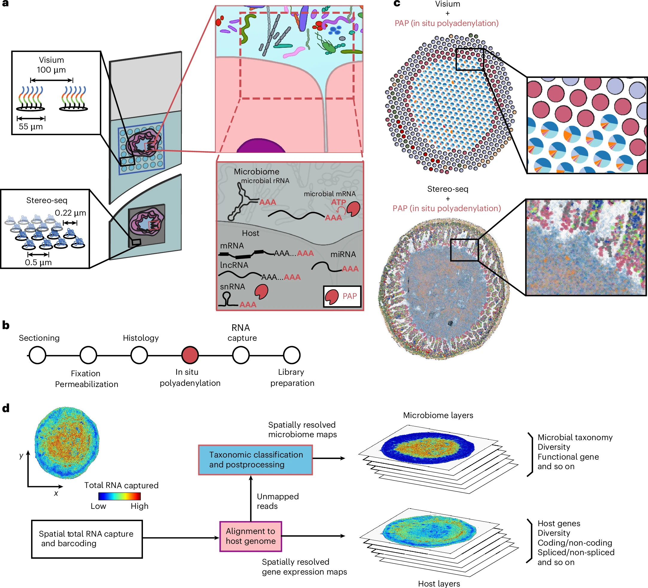
a, Overview of the experimental design. Array-based spatial RNA sequencing (at low or high spatial resolution) is combined with in situ polyadenylation via PAP. b, A schematic of the protocol for microbiome and host spatial total RNA sequencing. The standard steps of cryosectioning, fixation and histology are followed by enzymatic in situ polyadenylation, total RNA capture and sequencing library preparation. c, Representative data for the low (top) and high (bottom) resolution platforms. d, A schematic of bioinformatics workflow.
Using this strategy, the researchers were able to measure gene expression from both microbes and host cells while preserving their exact positions in the tissue. The technique achieves spatial resolution down to about one micrometer, allowing scientists to observe interactions occurring at very small distances between cells and bacteria.
When the method was applied to a mouse model of intestinal tumors, it revealed how microbial communities are organized throughout different regions of the intestine. The analysis showed that microbes often form tightly interacting clusters and that these interactions occur at very short distances.
The results also showed that the architecture of the host–microbiome interface changes in tumor tissue. These alterations may influence how microbes affect inflammation, immune responses, or tumor progression.
Importantly, the new workflow is compatible with widely used commercial spatial transcriptomics platforms. This means many laboratories can adopt the method without needing specialized equipment.
By enabling researchers to observe microbial and host gene activity at the same time and in the same location, this approach opens new opportunities for studying how the microbiome shapes health and disease. Understanding these interactions could help researchers uncover new mechanisms behind gastrointestinal disorders, cancer development, and microbiome driven immune responses.
Ntekas I, Takayasu L, McKellar D W, Grodner B, Holdener C, Schweitzer P, Park Y S, Sauthoff M, Shi Q, Brito I L, De Vlaminck I. (2026) Spatial transcriptomics maps host–gut microbiome biogeography at high resolution. Nature Microbiology [Epub ahead of print]. [article]
Related Posts






The human gut is home to trillions of microbes that constantly interact with each other and with the cells lining the intestine. These interactions influence digestion, immune responses, and even disease development. However, studying these relationships has been difficult because many techniques cannot show exactly where microbes and host cells are located relative to one another in tissue.
Researchers at Cornell University have developed a method that allows scientists to map these interactions at extremely high spatial resolution inside the gut. Their approach combines enzymatic modification of RNA molecules with spatial RNA sequencing, enabling the simultaneous analysis of bacterial and host gene activity directly within tissue samples.
One challenge in studying microbial activity in tissues is that bacterial RNA is often present in very small amounts compared with host RNA. To address this, the team introduced a step called enzymatic in situ polyadenylation, which adds poly(A) tails to bacterial RNA molecules. This modification allows bacterial transcripts to be captured more efficiently during sequencing, increasing sensitivity and making it easier to detect microbes that are present at low abundance.
In situ polyadenylation enables the capture of microbiome signals
with sequencing-based spatial transcriptomics platforms
a, Overview of the experimental design. Array-based spatial RNA sequencing (at low or high spatial resolution) is combined with in situ polyadenylation via PAP. b, A schematic of the protocol for microbiome and host spatial total RNA sequencing. The standard steps of cryosectioning, fixation and histology are followed by enzymatic in situ polyadenylation, total RNA capture and sequencing library preparation. c, Representative data for the low (top) and high (bottom) resolution platforms. d, A schematic of bioinformatics workflow.
Using this strategy, the researchers were able to measure gene expression from both microbes and host cells while preserving their exact positions in the tissue. The technique achieves spatial resolution down to about one micrometer, allowing scientists to observe interactions occurring at very small distances between cells and bacteria.
When the method was applied to a mouse model of intestinal tumors, it revealed how microbial communities are organized throughout different regions of the intestine. The analysis showed that microbes often form tightly interacting clusters and that these interactions occur at very short distances.
The results also showed that the architecture of the host–microbiome interface changes in tumor tissue. These alterations may influence how microbes affect inflammation, immune responses, or tumor progression.
Importantly, the new workflow is compatible with widely used commercial spatial transcriptomics platforms. This means many laboratories can adopt the method without needing specialized equipment.
By enabling researchers to observe microbial and host gene activity at the same time and in the same location, this approach opens new opportunities for studying how the microbiome shapes health and disease. Understanding these interactions could help researchers uncover new mechanisms behind gastrointestinal disorders, cancer development, and microbiome driven immune responses.
Ntekas I, Takayasu L, McKellar D W, Grodner B, Holdener C, Schweitzer P, Park Y S, Sauthoff M, Shi Q, Brito I L, De Vlaminck I. (2026) Spatial transcriptomics maps host–gut microbiome biogeography at high resolution. Nature Microbiology [Epub ahead of print]. [article]
Related Posts
Recent Posts
 Scientists identify the neuroblastoma cells that signal poor outcomes
 GARAGE improves rare cell detection in single-cell RNA sequencing analysis using AI
 Machine learning reveals how RNA splicing is regulated in individual cell types
 Illumina releases SpliceAI2 to help advance rare disease research
 Postdoctoral Fellow – Long-Read RNA Methods Development for Neurodegeneration
 Novel biosensor uses DNA ‘barcodes’ to uncover disease signals in cells
RNA-Seq Products & Services



Stay Connected