Researchers at EMBL Barcelona have quantitatively evaluated the risk of undetected artefacts in single-cell RNA sequencing experiments
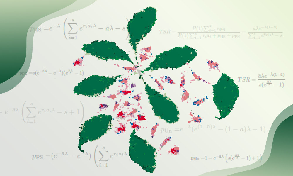
Fumio Nakaki combined theoretical modelling with experimental benchmarking and used publicly available single-cell datasets to assess how sample multiplexing influences multiplet detection. Credit: Fumio Nakaki/EMBL
Single-cell RNA sequencing (scRNA-seq) has revolutionised modern biology, allowing scientists to study the expression of genes of thousands of individual cells at the same time. This technique helps researchers identify different cell populations, study cellular heterogeneity, and understand gene expression dynamics in complex tissues.
In a recent study, researchers from EMBL Barcelona provide new insights into some of the technical limitations of the process, which could help scientists better address these risks while designing scRNA-seq experiments.
A common scRNA-seq method utilises a microfluidic device generating millions of ‘microdroplets’. Each cell is captured in a single microdroplet and then subjected to biochemical profiling. However, on random occasions, multiple cells are captured at the same time in a microdroplet and wrongly recognised as a single cell (called a ‘multiplet’).
This hidden artefact can distort data and mislead the interpretation of experiments. One way to reduce the risk of multiplets is ‘sample multiplexing’, a technique where samples are labelled with different molecular barcodes, and multiplets can be distinguished by the presence of multiple barcodes in the same microdroplet. Although this strategy has improved cost-efficiency and experimental design, it was unclear whether it still retained a risk of hidden multiplets.
In the new study, Fumio Nakaki, Postdoctoral Fellow at EMBL Barcelona’s Sharpe Group, has quantitatively evaluated the risk of having undetected (or ‘stealth’) multiplets during sample multiplexing, both in theory and in practice. His work has been published in BMC Genomics.
“Our goal is to provide researchers with a practical reference point,” said Nakaki. “Sample multiplexing is widely used, but its limitations are often overlooked. By clarifying how and when multiplets arise under multiplexed conditions, we hope this work will support better study design in experiments and data interpretation.”
To assess how sample multiplexing influences multiplet detection, Nakaki combined theoretical modelling with experimental benchmarking and used publicly available single-cell datasets. He derived mathematical expressions to estimate expected multiplet rates under various input conditions and validated these predictions against real data where individual samples had been uniquely barcoded.
Theoretical probability of the four types of multiplets in mx-scRNA-seq
and their presence in actual datasets

A Schematic illustration of the four types of multiplets possibly appearing in mx-scRNA-seq. Two-sample multiplex examples are shown. Labelled cells are shown in green or orange, depending on the sample labels, and unlabelled in white. The shapes represent the sample to which cells belong. In a sample multiplexing configuration, the multiplets are classified into four categories: Homogeneous stealth, partial stealth, multilabelled, and unlabelled multiplets, depending on combinations of cells in a single droplet. B The possible cell composition in a droplet containing at least one cell when two samples are multiplexed. The colour and shape of the cells are the same as (A). The probability of each category was calculated based on the combinatorics illustrated here. C An area chart exhibits the fraction of the four categories among multiplets in a hypothetical experiment, multiplexing five samples. is a parameter of the Poisson distribution, representing the average number of cells per droplet, closely correlated with a cell loading rate in practice. We assumed all samples had an equal labelling efficiency and were pooled with an equal proportion. Proportions were plotted against labelling efficiency when . D Proportions were plotted against the number of samples when a labelling efficiency was 0.9 and . E Proportions were plotted against when a labelling efficiency was 0.9 and 5 samples were multiplexed. FTSR is plotted against labelling efficiency under various sample numbers. is fixed at 0.3
By comparing different multiplexing configurations, Nakaki showed that while sample multiplexing helps identify some types of multiplets, at least one multiplet type still remains, especially in conditions where the sample labelling was insufficient or the choice of analysis tools was inadequate. Their analysis provides practical guidance on how to balance cell input, sample number, and desired specificity, highlighting that even with multiplexing, experimental parameters must be carefully optimised to maintain the integrity of single-cell data.
Source – EMBL


Researchers at EMBL Barcelona have quantitatively evaluated the risk of undetected artefacts in single-cell RNA sequencing experiments
Fumio Nakaki combined theoretical modelling with experimental benchmarking and used publicly available single-cell datasets to assess how sample multiplexing influences multiplet detection. Credit: Fumio Nakaki/EMBL
Single-cell RNA sequencing (scRNA-seq) has revolutionised modern biology, allowing scientists to study the expression of genes of thousands of individual cells at the same time. This technique helps researchers identify different cell populations, study cellular heterogeneity, and understand gene expression dynamics in complex tissues.
In a recent study, researchers from EMBL Barcelona provide new insights into some of the technical limitations of the process, which could help scientists better address these risks while designing scRNA-seq experiments.
A common scRNA-seq method utilises a microfluidic device generating millions of ‘microdroplets’. Each cell is captured in a single microdroplet and then subjected to biochemical profiling. However, on random occasions, multiple cells are captured at the same time in a microdroplet and wrongly recognised as a single cell (called a ‘multiplet’).
This hidden artefact can distort data and mislead the interpretation of experiments. One way to reduce the risk of multiplets is ‘sample multiplexing’, a technique where samples are labelled with different molecular barcodes, and multiplets can be distinguished by the presence of multiple barcodes in the same microdroplet. Although this strategy has improved cost-efficiency and experimental design, it was unclear whether it still retained a risk of hidden multiplets.
In the new study, Fumio Nakaki, Postdoctoral Fellow at EMBL Barcelona’s Sharpe Group, has quantitatively evaluated the risk of having undetected (or ‘stealth’) multiplets during sample multiplexing, both in theory and in practice. His work has been published in BMC Genomics.
To assess how sample multiplexing influences multiplet detection, Nakaki combined theoretical modelling with experimental benchmarking and used publicly available single-cell datasets. He derived mathematical expressions to estimate expected multiplet rates under various input conditions and validated these predictions against real data where individual samples had been uniquely barcoded.
Theoretical probability of the four types of multiplets in mx-scRNA-seq
and their presence in actual datasets
A Schematic illustration of the four types of multiplets possibly appearing in mx-scRNA-seq. Two-sample multiplex examples are shown. Labelled cells are shown in green or orange, depending on the sample labels, and unlabelled in white. The shapes represent the sample to which cells belong. In a sample multiplexing configuration, the multiplets are classified into four categories: Homogeneous stealth, partial stealth, multilabelled, and unlabelled multiplets, depending on combinations of cells in a single droplet. B The possible cell composition in a droplet containing at least one cell when two samples are multiplexed. The colour and shape of the cells are the same as (A). The probability of each category was calculated based on the combinatorics illustrated here. C An area chart exhibits the fraction of the four categories among multiplets in a hypothetical experiment, multiplexing five samples. is a parameter of the Poisson distribution, representing the average number of cells per droplet, closely correlated with a cell loading rate in practice. We assumed all samples had an equal labelling efficiency and were pooled with an equal proportion. Proportions were plotted against labelling efficiency when . D Proportions were plotted against the number of samples when a labelling efficiency was 0.9 and . E Proportions were plotted against when a labelling efficiency was 0.9 and 5 samples were multiplexed. FTSR is plotted against labelling efficiency under various sample numbers. is fixed at 0.3
By comparing different multiplexing configurations, Nakaki showed that while sample multiplexing helps identify some types of multiplets, at least one multiplet type still remains, especially in conditions where the sample labelling was insufficient or the choice of analysis tools was inadequate. Their analysis provides practical guidance on how to balance cell input, sample number, and desired specificity, highlighting that even with multiplexing, experimental parameters must be carefully optimised to maintain the integrity of single-cell data.
Source – EMBL
Nakaki F, Sharpe J. (2025) Probability of stealth multiplets in sample-multiplexing for droplet-based single-cell analysis. BMC Genomics 26(1): 686. [article]
Related Posts
Scientists identify the neuroblastoma cells that signal poor outcomes
Novel biosensor uses DNA ‘barcodes’ to uncover disease signals in cells
CellART extracts single-cell information from high-resolution spatial transcriptomics
Cell2space restores spatial context to single-cell RNA sequencing data
Urine test may improve bladder cancer diagnosis and guide treatment decisions
DeCoRE – a computational method to resolve RNA structural heterogeneity from RNA structure probing data by direct RNA sequencing
Tracing 700 million years of cell evolution
How should biological replicates be handled in long-read RNA sequencing?
RNA-Seq reveals a new way the developing heart controls its growth
The Human RNome Project Consortium seeks to publish first-ever complete sequence of the body’s RNA
scFlowReport brings multiple single-cell RNA sequencing analyses into one workflow
Long-read RNA sequencing reveals how neurons choose alternative RNA endings
DeOPUS improves cell-type estimates from bulk RNA sequencing
ResolVI improves accuracy in spatial transcriptomics analysis
RNA sequencing reveals transcriptional allele differences in prostate cancer stem cells
Unmasking “zombie cells” in aging tissue with an AI-powered barcode
RNA variant imaging for cancer diagnosis
Autism-risk mutations reveal two opposing patterns of brain gene activity
A real-data approach to evaluating RNA transcript analysis methods
Detection and sequencing of Ap2N-capped RNAs in human cells
Stay Connected
Submit a Post to the Blog
Recent Posts
Subscribe to the RNA-Seq Blog
RNA-Seq Products & Services