

Eosinophils are a type of white blood cell most people have heard of in the context of allergies or parasitic infections. But these cells actually play many roles in the body including helping control immune responses, supporting metabolism and aiding tissue repair. What hasn’t been clear until now is how eosinophils differ from one tissue to another and what makes them take on different roles in different environments. Researchers at Shanghai Jiao Tong University School of Medicine have created a detailed map showing how eosinophils vary depending on where they live in the body and how long they stay there.
To do this, the team looked at eosinophils from a variety of tissues in mice and analyzed both their RNA and surface proteins. They used advanced RNA sequencing to see which genes were active in individual cells, and combined that with detailed profiles of proteins on the cell surface. By tracing eosinophil development from bone marrow progenitors through different stages of life, the researchers found that tissue environment and residency time were major factors shaping eosinophil behavior.
Single-cell transcriptomics analysis identifies tissue-specific eosinophil states
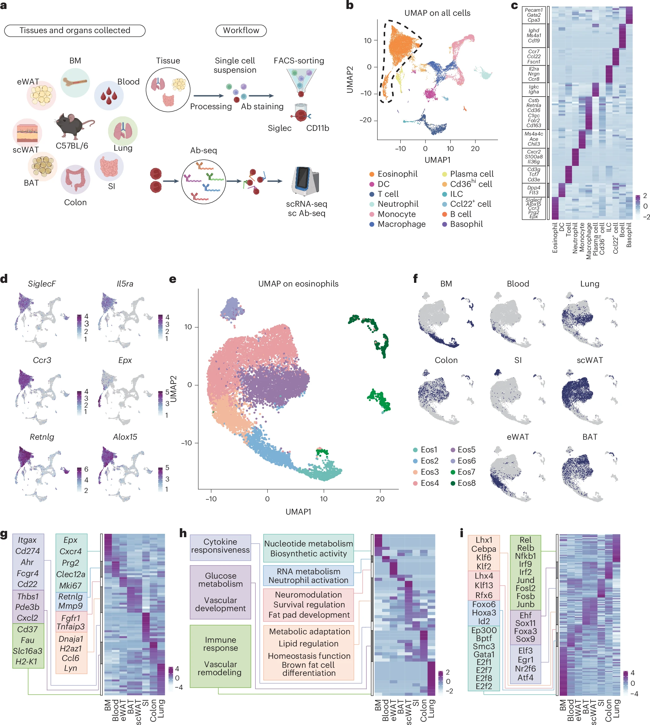
a, Schematic diagram of tissue processing, single-cell sequencing and antibody sequencing workflow. b, Integrated UMAP of all tissues with cell type annotations. DC, dendritic cell; ILC, innate lymphoid cell. c, Heatmap of DEGs across all cell populations. d, Expression of eosinophil marker genes. e, UMAP of eosinophil transcriptomes obtained from the BM, blood, lungs, colon, SI, scWAT, eWAT and BAT of C57BL/6 mice. f, Distribution of tissue origins projected on UMAP. g, Heatmap displaying tissue-specific DEG patterns. h, Tissue-originated pathway enrichment analysis shown by heatmap. i, TF regulatory network heatmap from SCENIC analysis across tissues (showing TF activation).
In the small intestine, where eosinophils tend to live a long time, cells became quite diverse, developing into distinct subtypes with different gene and protein signatures. In contrast, eosinophils in the lungs and colon, which have shorter lifespans, remained more similar to one another. The team also identified specific markers on the cell surface that track stages of maturation, helping distinguish newly formed eosinophils from those that have settled into a particular tissue.
This atlas gives scientists a unified view of how tissue cues and life history drive eosinophil specialization. These insights make it easier to identify eosinophil states in health, such as maintaining normal tissue function, and in disease, where eosinophils can contribute to inflammation. Having a molecular “toolkit” to resolve eosinophil states will help researchers and clinicians better understand and potentially target these cells in conditions like asthma, inflammatory bowel disease and beyond.
Li Hu Y, Wu Lianfu, Qu Sihao, Kong Wan Ting, Yu Xiaotong, Xu Jiaqian, Xu Ziyi, He Jialing, Wang Chong, Liu Zhaoyuan, Lu Liming, Ng Lai Guan, Blériot Camille, Su Bing, Ginhoux Florent, Li Ziyi, Chakarov Svetoslav (2026) Temporal and spatial atlas of eosinophil specialization across tissues. Nature Immunology [Epub ahead of print]. [article]
Related Posts






Eosinophils are a type of white blood cell most people have heard of in the context of allergies or parasitic infections. But these cells actually play many roles in the body including helping control immune responses, supporting metabolism and aiding tissue repair. What hasn’t been clear until now is how eosinophils differ from one tissue to another and what makes them take on different roles in different environments. Researchers at Shanghai Jiao Tong University School of Medicine have created a detailed map showing how eosinophils vary depending on where they live in the body and how long they stay there.
To do this, the team looked at eosinophils from a variety of tissues in mice and analyzed both their RNA and surface proteins. They used advanced RNA sequencing to see which genes were active in individual cells, and combined that with detailed profiles of proteins on the cell surface. By tracing eosinophil development from bone marrow progenitors through different stages of life, the researchers found that tissue environment and residency time were major factors shaping eosinophil behavior.
Single-cell transcriptomics analysis identifies tissue-specific eosinophil states
a, Schematic diagram of tissue processing, single-cell sequencing and antibody sequencing workflow. b, Integrated UMAP of all tissues with cell type annotations. DC, dendritic cell; ILC, innate lymphoid cell. c, Heatmap of DEGs across all cell populations. d, Expression of eosinophil marker genes. e, UMAP of eosinophil transcriptomes obtained from the BM, blood, lungs, colon, SI, scWAT, eWAT and BAT of C57BL/6 mice. f, Distribution of tissue origins projected on UMAP. g, Heatmap displaying tissue-specific DEG patterns. h, Tissue-originated pathway enrichment analysis shown by heatmap. i, TF regulatory network heatmap from SCENIC analysis across tissues (showing TF activation).
In the small intestine, where eosinophils tend to live a long time, cells became quite diverse, developing into distinct subtypes with different gene and protein signatures. In contrast, eosinophils in the lungs and colon, which have shorter lifespans, remained more similar to one another. The team also identified specific markers on the cell surface that track stages of maturation, helping distinguish newly formed eosinophils from those that have settled into a particular tissue.
This atlas gives scientists a unified view of how tissue cues and life history drive eosinophil specialization. These insights make it easier to identify eosinophil states in health, such as maintaining normal tissue function, and in disease, where eosinophils can contribute to inflammation. Having a molecular “toolkit” to resolve eosinophil states will help researchers and clinicians better understand and potentially target these cells in conditions like asthma, inflammatory bowel disease and beyond.
Li Hu Y, Wu Lianfu, Qu Sihao, Kong Wan Ting, Yu Xiaotong, Xu Jiaqian, Xu Ziyi, He Jialing, Wang Chong, Liu Zhaoyuan, Lu Liming, Ng Lai Guan, Blériot Camille, Su Bing, Ginhoux Florent, Li Ziyi, Chakarov Svetoslav (2026) Temporal and spatial atlas of eosinophil specialization across tissues. Nature Immunology [Epub ahead of print]. [article]
Related Posts
Recent Posts
 Scientists identify the neuroblastoma cells that signal poor outcomes
 GARAGE improves rare cell detection in single-cell RNA sequencing analysis using AI
 Machine learning reveals how RNA splicing is regulated in individual cell types
 Illumina releases SpliceAI2 to help advance rare disease research
 Postdoctoral Fellow – Long-Read RNA Methods Development for Neurodegeneration
 Novel biosensor uses DNA ‘barcodes’ to uncover disease signals in cells
RNA-Seq Products & Services



Stay Connected