

Herpes Simplex Virus 1, or HSV-1, is a common virus that many people carry, often causing cold sores. However, in some cases, HSV-1 can infect the brain, leading to serious conditions like encephalitis, which can cause brain inflammation and long-term neurological problems. Researchers at Aarhus University have used advanced RNA sequencing technologies to better understand how the brain fights this infection and how the immune response may also contribute to brain damage.
Model for the antiviral and inflammatory activities in the HSV-1-infected mouse brain
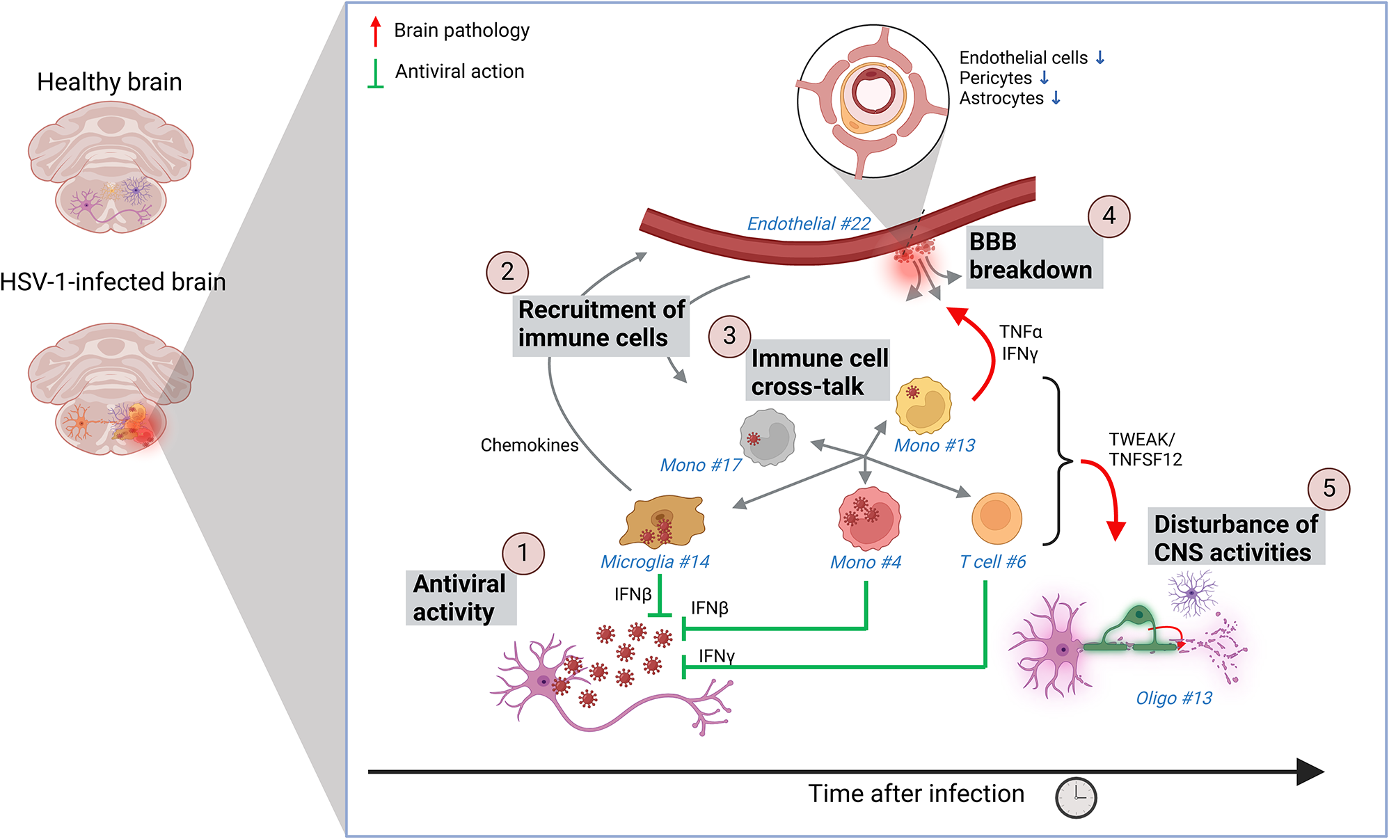
HSV-1 infection induces early expression of an innate antiviral IFN response, mainly driven by a microglia subpopulation, that is characteristic for virus infections (virus-activated microglia, VAM). This is paralleled by expression of cytokines and chemokines, which trigger recruitment of immune cells, mainly monocytes. The monocytes include an array of subpopulations, and engage in extensive cross-talk with other immune cell populations, thus amplifying their activities. Through the accompanying inflammatory response, microglia, monocytes, and T cells cross-talks with brain structures and functions, including the BBB and the oligodendrocyte-neuron unit. These interactions involve numerous pathways, including TNFα, IFNγ, and TNFSF12/TWEAK and lead to loss of BBB integrity and neuro-physiological functions. Eventually, this can cause acute disease, or long-term sequelae
Using two cutting-edge methods called single-cell RNA sequencing and GeoMx spatial transcriptomics, the researchers studied infected mouse brain stems in detail. These techniques allowed them to look at individual cells and how they communicate during infection. They discovered that shortly after HSV-1 infects the brain, microglia, the brain’s resident immune cells, increase in number and become highly active. These microglia produce antiviral molecules and chemical signals called chemokines that call other immune cells, such as monocytes and T cells, to the site of infection.
Monocytes, a type of immune cell that normally circulates in the blood, enter the brain and show an even broader range of activity than microglia. These cells act like coordinators, interacting with many different cell types to mount an immune defense. Unfortunately, the infection also damages the blood-brain barrier, which normally protects the brain from harmful substances in the blood. This damage allows more immune cells to enter the brain but also leads to disruption of normal brain function.
The study found that cytokines, small signaling proteins released by microglia, monocytes, and T cells, change the behavior of brain cells such as endothelial cells (which line blood vessels) and oligodendrocytes (which help insulate nerve fibers). These changes can disturb normal brain activity and contribute to disease symptoms.
An exciting discovery was the identification of a unique subpopulation of microglia that appears only during viral brain infections, with strong antiviral and inflammatory activity. This finding suggests that microglia respond differently to viruses than to other types of brain injuries or diseases, which may open up new paths for targeted treatments.
Overall, these findings improve our understanding of how the immune system and brain cells interact during HSV-1 infection. This knowledge could help scientists develop better therapies that not only fight the virus but also reduce harmful inflammation and protect brain function.
Tryggvason E, Henze A, Eriksson J, Gatti D, Davies C, Malm T, Khademi M, Brundin L, Ingelsson M, Nilsson P. (2025) Spatial and single-cell transcriptomic profiling reveals virus-induced microglia and monocyte interactions driving brain pathology in HSV-1 infection. Molecular Therapy [Online ahead of print]. [article]
Related Posts






Herpes Simplex Virus 1, or HSV-1, is a common virus that many people carry, often causing cold sores. However, in some cases, HSV-1 can infect the brain, leading to serious conditions like encephalitis, which can cause brain inflammation and long-term neurological problems. Researchers at Aarhus University have used advanced RNA sequencing technologies to better understand how the brain fights this infection and how the immune response may also contribute to brain damage.
Model for the antiviral and inflammatory activities in the HSV-1-infected mouse brain
HSV-1 infection induces early expression of an innate antiviral IFN response, mainly driven by a microglia subpopulation, that is characteristic for virus infections (virus-activated microglia, VAM). This is paralleled by expression of cytokines and chemokines, which trigger recruitment of immune cells, mainly monocytes. The monocytes include an array of subpopulations, and engage in extensive cross-talk with other immune cell populations, thus amplifying their activities. Through the accompanying inflammatory response, microglia, monocytes, and T cells cross-talks with brain structures and functions, including the BBB and the oligodendrocyte-neuron unit. These interactions involve numerous pathways, including TNFα, IFNγ, and TNFSF12/TWEAK and lead to loss of BBB integrity and neuro-physiological functions. Eventually, this can cause acute disease, or long-term sequelae
Using two cutting-edge methods called single-cell RNA sequencing and GeoMx spatial transcriptomics, the researchers studied infected mouse brain stems in detail. These techniques allowed them to look at individual cells and how they communicate during infection. They discovered that shortly after HSV-1 infects the brain, microglia, the brain’s resident immune cells, increase in number and become highly active. These microglia produce antiviral molecules and chemical signals called chemokines that call other immune cells, such as monocytes and T cells, to the site of infection.
Monocytes, a type of immune cell that normally circulates in the blood, enter the brain and show an even broader range of activity than microglia. These cells act like coordinators, interacting with many different cell types to mount an immune defense. Unfortunately, the infection also damages the blood-brain barrier, which normally protects the brain from harmful substances in the blood. This damage allows more immune cells to enter the brain but also leads to disruption of normal brain function.
The study found that cytokines, small signaling proteins released by microglia, monocytes, and T cells, change the behavior of brain cells such as endothelial cells (which line blood vessels) and oligodendrocytes (which help insulate nerve fibers). These changes can disturb normal brain activity and contribute to disease symptoms.
An exciting discovery was the identification of a unique subpopulation of microglia that appears only during viral brain infections, with strong antiviral and inflammatory activity. This finding suggests that microglia respond differently to viruses than to other types of brain injuries or diseases, which may open up new paths for targeted treatments.
Overall, these findings improve our understanding of how the immune system and brain cells interact during HSV-1 infection. This knowledge could help scientists develop better therapies that not only fight the virus but also reduce harmful inflammation and protect brain function.
Tryggvason E, Henze A, Eriksson J, Gatti D, Davies C, Malm T, Khademi M, Brundin L, Ingelsson M, Nilsson P. (2025) Spatial and single-cell transcriptomic profiling reveals virus-induced microglia and monocyte interactions driving brain pathology in HSV-1 infection. Molecular Therapy [Online ahead of print]. [article]
Related Posts
Recent Posts
 RNAmel simplifies RNA sequencing data analysis with a reproducible, integrated platform
 Heart failure reshapes bone marrow to create inflammation-related immune memory
 Overlooked DNA regions surrounding ‘microexons’ hold missing pieces of autism’s genetic puzzle
 RiboBA improves detection of hidden protein-coding regions using ribosome profiling
 An anti-senescence gene may fight spinal disc deterioration
 Bulk RNA-seq Analysis: A Complete Beginner’s Guide
RNA-Seq Products & Services


Stay Connected