

Researchers from the Centre for Genomic Regulation (CRG) in Barcelona explore how scientists are using advanced sequencing tools to study chemical changes on RNA. These RNA modifications can affect how cells function, but detecting them accurately has been a major challenge.
Nanopore direct RNA sequencing has opened new doors by allowing researchers to read RNA molecules in their natural form. Unlike older methods that required copying RNA into DNA, nanopore sequencing measures the electrical signal from each nucleotide directly. Because chemical modifications change this signal, they can theoretically be detected as the molecule passes through the nanopore.
Comparison of m6A stoichiometry predictions using different m6A base-calling models
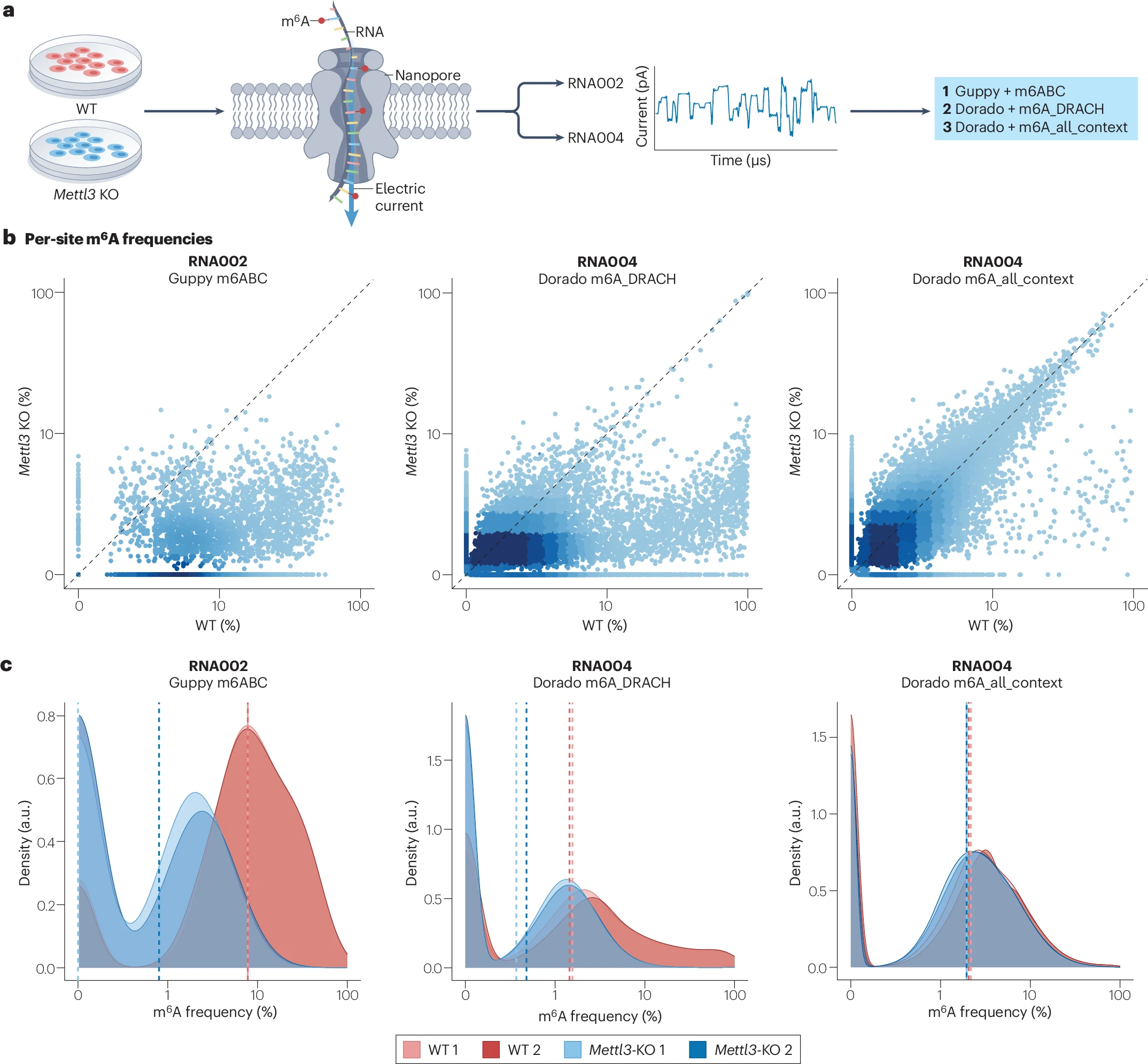
a, An overview of the comparative analysis. The RNA from wild-type (WT) and from inducible Mettl3-knockout (KO) mouse embryonic stem cells (mESCs) was sequenced in two replicates, with both SQK-RNA002 and SQK-RNA004 chemistries. The raw sequencing output was processed with m6ABasecaller (m6ABC) for SQK-RNA002 and with dorado m6A_DRACH and m6A_all_context models for SQK-RNA004. b, Scatterplots of per-site N6-methyladenosine (m6A) frequencies (mean of two replicates) of WT versus Mettl3-KO mESC samples, calculated with the three base-calling models. Both axes are in logarithmic scale. A gradient from light to dark blue depicts the increase in data points density in the plot. The diagonal dashed line indicates the x = y line in frequencies. Only sites with 25 reads or more of coverage in all samples and frequency greater than 0% in at least one sample for each method were included in the analysis. c, A density plot distribution of the m6A frequency in WT and Mettl3-KO replicates. The vertical dashed lines indicate the median m6A modification frequency in each sample. The x axis is in logarithmic scale. Only sites with 25 reads or more of coverage in all samples for each method were included in the analysis.
To make this easier, researchers have developed special base calling models. These are software tools trained to recognize modified RNA bases such as:
- m6A
- inosine (I)
- pseudouridine (Ψ)
- m5C
These models claim to detect modifications at single molecule resolution, which could dramatically boost our understanding of the epitranscriptome, the layer of regulation that involves chemical marks on RNA.
However, the researchers highlight several important limitations that scientists must keep in mind. The performance of these models is still unclear, and each one can behave differently. Some may mistake one modification for another, thresholds for calling modifications are not standardized, and false positive rates vary widely. When the researchers tested three different models on identical RNA samples, they saw over 20-fold differences in the number of predicted m6A sites.
This huge variation shows why caution is essential. Without proper validation and consistent analysis pipelines, results could be misinterpreted and lead researchers in the wrong direction. As these tools become more popular, understanding their weaknesses will be just as important as celebrating their capabilities.
Overall, the researchers emphasize the promise of nanopore RNA sequencing while reminding the scientific community that accuracy, standardization, and validation remain crucial as we move deeper into single molecule epitranscriptomics.
Cruciani S, Novoa E.M. (2025) The new era of single molecule RNA modification detection through nanopore base calling models. Nature Reviews Molecular Cell Biology [Online ahead of print]. [article]
Related Posts


Researchers from the Centre for Genomic Regulation (CRG) in Barcelona explore how scientists are using advanced sequencing tools to study chemical changes on RNA. These RNA modifications can affect how cells function, but detecting them accurately has been a major challenge.
Nanopore direct RNA sequencing has opened new doors by allowing researchers to read RNA molecules in their natural form. Unlike older methods that required copying RNA into DNA, nanopore sequencing measures the electrical signal from each nucleotide directly. Because chemical modifications change this signal, they can theoretically be detected as the molecule passes through the nanopore.
Comparison of m6A stoichiometry predictions using different m6A base-calling models
a, An overview of the comparative analysis. The RNA from wild-type (WT) and from inducible Mettl3-knockout (KO) mouse embryonic stem cells (mESCs) was sequenced in two replicates, with both SQK-RNA002 and SQK-RNA004 chemistries. The raw sequencing output was processed with m6ABasecaller (m6ABC) for SQK-RNA002 and with dorado m6A_DRACH and m6A_all_context models for SQK-RNA004. b, Scatterplots of per-site N6-methyladenosine (m6A) frequencies (mean of two replicates) of WT versus Mettl3-KO mESC samples, calculated with the three base-calling models. Both axes are in logarithmic scale. A gradient from light to dark blue depicts the increase in data points density in the plot. The diagonal dashed line indicates the x = y line in frequencies. Only sites with 25 reads or more of coverage in all samples and frequency greater than 0% in at least one sample for each method were included in the analysis. c, A density plot distribution of the m6A frequency in WT and Mettl3-KO replicates. The vertical dashed lines indicate the median m6A modification frequency in each sample. The x axis is in logarithmic scale. Only sites with 25 reads or more of coverage in all samples for each method were included in the analysis.
To make this easier, researchers have developed special base calling models. These are software tools trained to recognize modified RNA bases such as:
- m6A
- inosine (I)
- pseudouridine (Ψ)
- m5C
These models claim to detect modifications at single molecule resolution, which could dramatically boost our understanding of the epitranscriptome, the layer of regulation that involves chemical marks on RNA.
However, the researchers highlight several important limitations that scientists must keep in mind. The performance of these models is still unclear, and each one can behave differently. Some may mistake one modification for another, thresholds for calling modifications are not standardized, and false positive rates vary widely. When the researchers tested three different models on identical RNA samples, they saw over 20-fold differences in the number of predicted m6A sites.
This huge variation shows why caution is essential. Without proper validation and consistent analysis pipelines, results could be misinterpreted and lead researchers in the wrong direction. As these tools become more popular, understanding their weaknesses will be just as important as celebrating their capabilities.
Overall, the researchers emphasize the promise of nanopore RNA sequencing while reminding the scientific community that accuracy, standardization, and validation remain crucial as we move deeper into single molecule epitranscriptomics.
Cruciani S, Novoa E.M. (2025) The new era of single molecule RNA modification detection through nanopore base calling models. Nature Reviews Molecular Cell Biology [Online ahead of print]. [article]
Related Posts
Recent Posts
 Scientists identify the neuroblastoma cells that signal poor outcomes
 GARAGE improves rare cell detection in single-cell RNA sequencing analysis using AI
 Machine learning reveals how RNA splicing is regulated in individual cell types
 Illumina releases SpliceAI2 to help advance rare disease research
 Postdoctoral Fellow – Long-Read RNA Methods Development for Neurodegeneration
 Novel biosensor uses DNA ‘barcodes’ to uncover disease signals in cells
RNA-Seq Products & Services


Stay Connected