

Single-cell sequencing is a powerful tool in biology, allowing scientists to study individual cells in great detail. This method has opened up new ways to understand how different cells in a tissue behave, interact, and contribute to diseases like cancer. One of the challenges with single-cell analysis, though, is that many methods are either costly or require specific protocols for different types of experiments. This has limited their use in clinical settings where high throughput, scalability, and efficiency are crucial.
A new approach developed at the Beijing Institute of Genomics, called UDA-seq, aims to solve these problems by introducing a universal workflow for single-cell sequencing. The UDA-seq method improves on existing techniques by incorporating a post-indexing step that enhances throughput. This step allows researchers to adapt existing single-cell sequencing methods for a variety of applications, making the process more automated, scalable, and accessible.
The workflow of UDA-seq and its performance across diverse modalities and samples
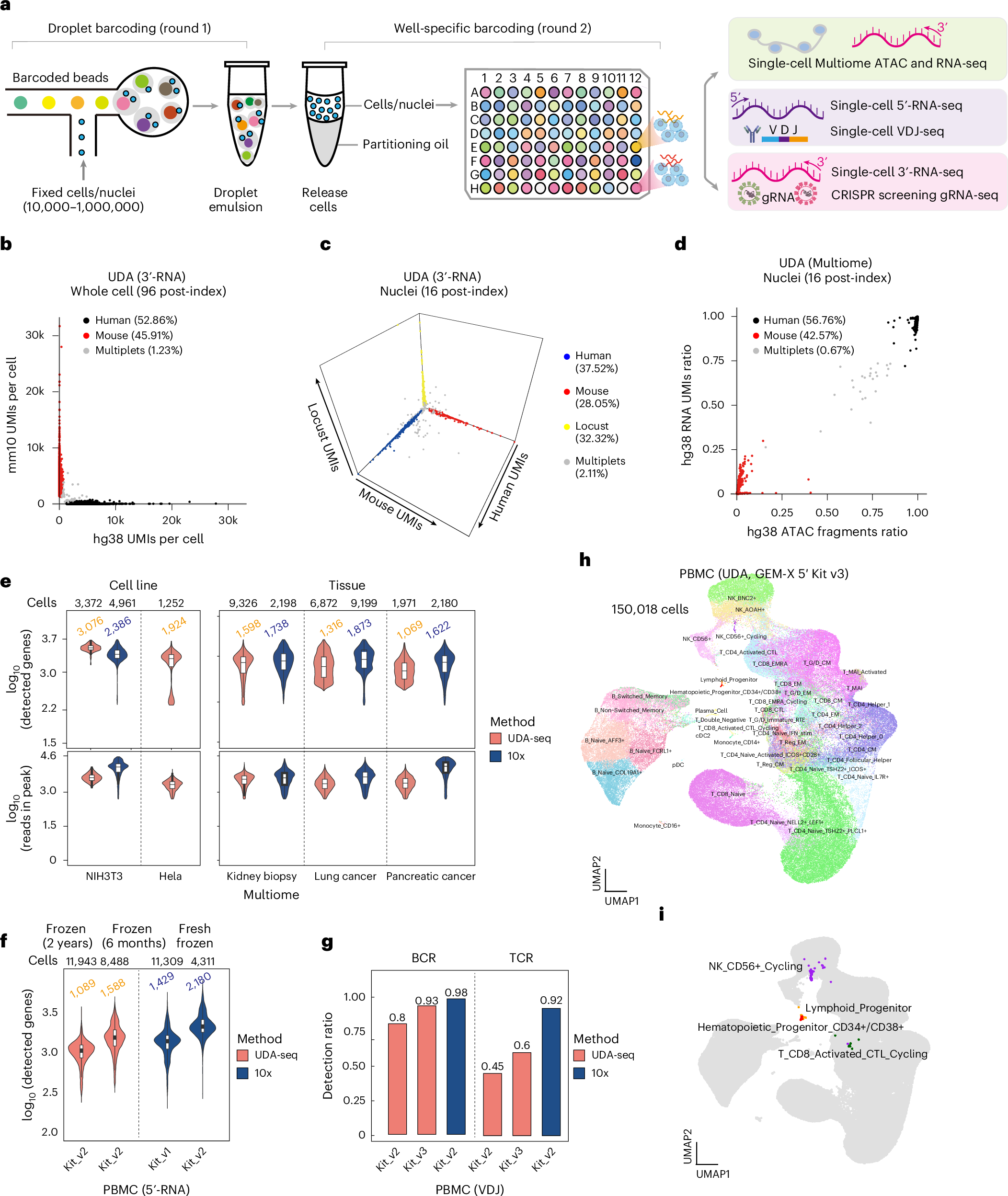
a, The UDA-seq workflow involves two rounds of barcoding. b–d, Species-mixing experiments by using single-cell 3′-RNA UDA-seq with 96-well post-indexing (b), single-nucleus 3′-RNA UDA-seq with 16 well post-indexing (c) and single-nucleus scMultiome UDA-seq with 16 well post-indexing (d). e,f, Violin plots showing the distribution of genes (RNA) and peak reads (ATAC) detected by UDA-seq scMultiome and 10x Genomics (e) as well as UDA-seq sc5′-RNA (f). Box plots show the interquartile range with the median marked. The whiskers extend up to 1.5 times the interquartile range; the outliers are not displayed. The median number of genes detected is shown at the top of each violin plot. g, A comparison between UDA and 10x Genomics for scVDJ-seq. The T-cell receptor (TCR) and B-cell receptor (BCR) detection ratio for UDA with Next GEM (Kit_v2) and GEM-X (Kit_v3), and the 10x Next GEM. h, The ultrahigh-throughput 5′-scRNA UDA-seq analysis of over 150,000 cells from a single donor, identifying 44 distinct cell types. The UMAP plot is color-coded to represent these cell types. j, Rare cell types, identified with cell type labels.
What Makes UDA-seq Different?
Traditional single-cell sequencing methods often require specialized protocols for each analysis. For example, if you want to study the gene expression of RNA alongside other cellular features like DNA mutations or chromatin modifications, existing methods would need to be tailored for each of those. UDA-seq simplifies this by offering a universal approach that can handle multiple types of analyses, such as RNA and DNA sequencing, or RNA and chromatin analysis, all in one experiment.
The method was tested across different tissues and cell types, which demonstrated its versatility. The researchers successfully combined common single-cell analyses like RNA and VDJ sequencing, RNA and chromatin profiling, and RNA alongside CRISPR-based perturbations (a tool used to edit genes) in a single workflow. This flexibility is a major advantage, as it reduces the complexity and costs associated with traditional single-cell analyses that require separate experiments for each type of data.
Scalability for Clinical Applications
One of the most exciting aspects of UDA-seq is its potential for clinical use. In a study using frozen clinical biopsy specimens, UDA-seq helped the researchers generate over 100,000 high-quality single-cell datasets. This was all done in a single experiment using a single-channel droplet microfluidics setup. Droplet microfluidics is a technique that allows researchers to analyze many individual cells at once by encapsulating each cell in a small droplet, making the process much more efficient.
This high throughput capability means that UDA-seq could be applied in clinical settings, where large-scale cell analysis is needed for understanding diseases like cancer. For instance, researchers can now identify rare cell subpopulations that are linked to specific clinical outcomes, such as the development or progression of cancer. By analyzing the genetic makeup and characteristics of these cells, UDA-seq helps researchers explore how certain cells in cancer might be more vulnerable to treatment, offering new insights for personalized medicine.
Conclusion
UDA-seq represents a significant advancement in single-cell sequencing technology. By offering a universal, scalable, and automated workflow, it has the potential to make single-cell analysis more accessible, especially in clinical and cancer research. This approach allows researchers to study complex cell populations and their behaviors in ways that were previously too costly or complicated, bringing us closer to personalized treatments and a deeper understanding of disease mechanisms.
Li Y, Huang Z, Xu L et al. (2025) UDA-seq: universal droplet microfluidics-based combinatorial indexing for massive-scale multimodal single-cell sequencing. Nat Methods [Epub ahead of print]. [article]
Related Posts




Single-cell sequencing is a powerful tool in biology, allowing scientists to study individual cells in great detail. This method has opened up new ways to understand how different cells in a tissue behave, interact, and contribute to diseases like cancer. One of the challenges with single-cell analysis, though, is that many methods are either costly or require specific protocols for different types of experiments. This has limited their use in clinical settings where high throughput, scalability, and efficiency are crucial.
A new approach developed at the Beijing Institute of Genomics, called UDA-seq, aims to solve these problems by introducing a universal workflow for single-cell sequencing. The UDA-seq method improves on existing techniques by incorporating a post-indexing step that enhances throughput. This step allows researchers to adapt existing single-cell sequencing methods for a variety of applications, making the process more automated, scalable, and accessible.
The workflow of UDA-seq and its performance across diverse modalities and samples
a, The UDA-seq workflow involves two rounds of barcoding. b–d, Species-mixing experiments by using single-cell 3′-RNA UDA-seq with 96-well post-indexing (b), single-nucleus 3′-RNA UDA-seq with 16 well post-indexing (c) and single-nucleus scMultiome UDA-seq with 16 well post-indexing (d). e,f, Violin plots showing the distribution of genes (RNA) and peak reads (ATAC) detected by UDA-seq scMultiome and 10x Genomics (e) as well as UDA-seq sc5′-RNA (f). Box plots show the interquartile range with the median marked. The whiskers extend up to 1.5 times the interquartile range; the outliers are not displayed. The median number of genes detected is shown at the top of each violin plot. g, A comparison between UDA and 10x Genomics for scVDJ-seq. The T-cell receptor (TCR) and B-cell receptor (BCR) detection ratio for UDA with Next GEM (Kit_v2) and GEM-X (Kit_v3), and the 10x Next GEM. h, The ultrahigh-throughput 5′-scRNA UDA-seq analysis of over 150,000 cells from a single donor, identifying 44 distinct cell types. The UMAP plot is color-coded to represent these cell types. j, Rare cell types, identified with cell type labels.
What Makes UDA-seq Different?
Traditional single-cell sequencing methods often require specialized protocols for each analysis. For example, if you want to study the gene expression of RNA alongside other cellular features like DNA mutations or chromatin modifications, existing methods would need to be tailored for each of those. UDA-seq simplifies this by offering a universal approach that can handle multiple types of analyses, such as RNA and DNA sequencing, or RNA and chromatin analysis, all in one experiment.
The method was tested across different tissues and cell types, which demonstrated its versatility. The researchers successfully combined common single-cell analyses like RNA and VDJ sequencing, RNA and chromatin profiling, and RNA alongside CRISPR-based perturbations (a tool used to edit genes) in a single workflow. This flexibility is a major advantage, as it reduces the complexity and costs associated with traditional single-cell analyses that require separate experiments for each type of data.
Scalability for Clinical Applications
One of the most exciting aspects of UDA-seq is its potential for clinical use. In a study using frozen clinical biopsy specimens, UDA-seq helped the researchers generate over 100,000 high-quality single-cell datasets. This was all done in a single experiment using a single-channel droplet microfluidics setup. Droplet microfluidics is a technique that allows researchers to analyze many individual cells at once by encapsulating each cell in a small droplet, making the process much more efficient.
This high throughput capability means that UDA-seq could be applied in clinical settings, where large-scale cell analysis is needed for understanding diseases like cancer. For instance, researchers can now identify rare cell subpopulations that are linked to specific clinical outcomes, such as the development or progression of cancer. By analyzing the genetic makeup and characteristics of these cells, UDA-seq helps researchers explore how certain cells in cancer might be more vulnerable to treatment, offering new insights for personalized medicine.
Conclusion
UDA-seq represents a significant advancement in single-cell sequencing technology. By offering a universal, scalable, and automated workflow, it has the potential to make single-cell analysis more accessible, especially in clinical and cancer research. This approach allows researchers to study complex cell populations and their behaviors in ways that were previously too costly or complicated, bringing us closer to personalized treatments and a deeper understanding of disease mechanisms.
Li Y, Huang Z, Xu L et al. (2025) UDA-seq: universal droplet microfluidics-based combinatorial indexing for massive-scale multimodal single-cell sequencing. Nat Methods [Epub ahead of print]. [article]
Related Posts
Recent Posts
 Machine learning reveals how RNA splicing is regulated in individual cell types
 Illumina releases SpliceAI2 to help advance rare disease research
 Postdoctoral Fellow – Long-Read RNA Methods Development for Neurodegeneration
 Novel biosensor uses DNA ‘barcodes’ to uncover disease signals in cells
 GP-DHT uses single-cell RNA sequencing to map gene regulatory networks
 Boehringer taps Envisagenics’ AI to mine RNA splicing for cancer targets
RNA-Seq Products & Services


Stay Connected