Cornell researchers have uncovered the genetic triggers that cause male and female bovine embryos to develop differently, as early as seven to eight days after fertilization. The breakthrough in basic science has implications for human health, such as drug development and in vitro fertilization, and for bovine health and dairy industry sustainability.
Scientists have known since the 1990s that male embryos of multiple mammalian species, including humans, grow faster than female embryos, but until now, the underlying reasons were unclear.
In a new paper, published Aug. 27 in Cell & Bioscience, Cornell scientists grew bovine embryos in petri dishes then analyzed their genetic sex and RNA sequencing, which shows how genes are being expressed. They discovered significant sex differences in gene regulation: Male embryos prioritized genes associated with energy metabolism, causing them to grow faster than their female counterparts. Female embryos emphasized genes associated with sex differentiation, gonad development and inflammatory pathways that are important for future development.
Overview of embryo sex determination
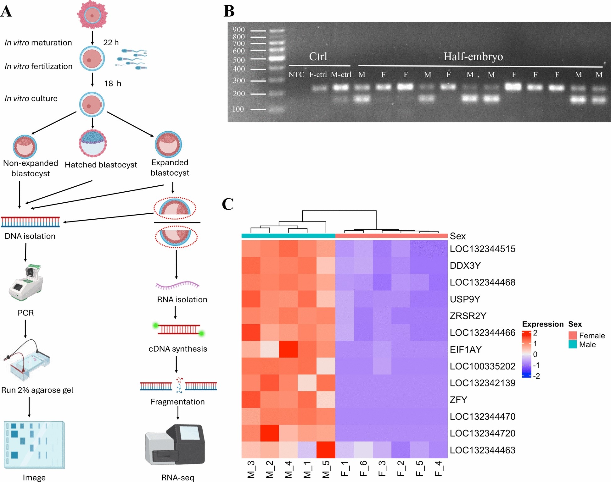
A Schematic overview of the experimental setup. Expanded blastocysts were cut into two halves evenly, with each half containing inner cell mass (ICM), trophectoderm (TE), and zona pellucida. One half was used for RNA-seq, and the other half was used for PCR-based sex determination. B Agarose gel of amplified gDNA of half blastocysts. Each lane is one control sample or a half-cut embryo; NTC: no-template control; F-ctrl: gDNA from bovine mammary alveolar cells (MAC-T cells); M-ctrl: gDNA from bovine testis tissues. Identification of the autosomal amplicon (217 bp) located above and the Y-specific amplicon (143 bp) located below. C Heatmap of all male-specific DEGs located on Y-chromosome in all RNA-seq samples. Each column represented an individual RNA-seq sample, and the hierarchical clustering along the X axis represented sample similarity based on the selected Y-linked gene expression. The color scale in the heatmap represents the z-score of normalized gene expression
Understanding these fundamental sex differences at the genomic and molecular levels is critically important to improving in vitro fertilization (IVF) success in humans and cows, and in developing treatments that will work for both men and women, said Jingyue “Ellie” Duan, assistant professor of functional genomics in the College of Agriculture and Life Sciences and paper co-author. Co-first authors are Meihong Shi, a postdoctoral associate, and Guangsheng Li, a Ph.D. candidate, both in Duan’s lab.
“Sex difference has been a factor ignored in a lot of studies and clinical trials. Until very recently, most mouse studies have used exclusively male mice. And most drug discovery is done with male mice,” Duan said. “And yet, we see that onset and occurrence of many diseases are different in men and women: Alzheimer’s, autoimmune diseases, heart disease. In this basic study, we found that male and female embryos are different, even at this very early stage, on the basis of genome regulation.”
At this early stage, sex-associated hormones like estrogen and testosterone – which contribute to average size differences between male and female adults – have not yet come into play. Advances in genome sequencing technology enabled researchers to revisit the question at a genome-wide level, Duan said.
“This could imply that there’s an intrinsic factor in our genome contributing to the sex-based differences we see, such as sex chromosomes (XX versus XY) or sex-linked genes, not just from hormonal change or environmental factors,” Duan said. “We’re born with this sex-specific genetic regulation that is contributing very differently to cellular behavior, disease onset and immune system development, and continues through life all the way to health and aging. That’s a very important message for people in the clinical field and people developing drugs to understand.”
Bovines are an effective animal model for human-health studies, but bovine research is also important for its role in supporting food supplies and sustainable agriculture. Understanding and optimizing bovine reproduction is especially important for the dairy industry, which relies on cattle reproduction through IVF to maintain milk supplies.
Duan’s lab collaborated in this research with the lab of Soon Hon Cheong, Ph.D. ’12, associate professor of clinical sciences in the College of Veterinary Medicine. The project would not have been possible without collaboration between Duan’s genomics-focused lab and Cheong’s group, which studies reproductive medicine and assisted reproductive techniques, Duan said.
Future research, already underway in both labs, will expand this work by studying sex differences in embryos from fertilization through day eight after fertilization.
Source – Cornell University


Cornell researchers have uncovered the genetic triggers that cause male and female bovine embryos to develop differently, as early as seven to eight days after fertilization. The breakthrough in basic science has implications for human health, such as drug development and in vitro fertilization, and for bovine health and dairy industry sustainability.
Scientists have known since the 1990s that male embryos of multiple mammalian species, including humans, grow faster than female embryos, but until now, the underlying reasons were unclear.
In a new paper, published Aug. 27 in Cell & Bioscience, Cornell scientists grew bovine embryos in petri dishes then analyzed their genetic sex and RNA sequencing, which shows how genes are being expressed. They discovered significant sex differences in gene regulation: Male embryos prioritized genes associated with energy metabolism, causing them to grow faster than their female counterparts. Female embryos emphasized genes associated with sex differentiation, gonad development and inflammatory pathways that are important for future development.
Overview of embryo sex determination
A Schematic overview of the experimental setup. Expanded blastocysts were cut into two halves evenly, with each half containing inner cell mass (ICM), trophectoderm (TE), and zona pellucida. One half was used for RNA-seq, and the other half was used for PCR-based sex determination. B Agarose gel of amplified gDNA of half blastocysts. Each lane is one control sample or a half-cut embryo; NTC: no-template control; F-ctrl: gDNA from bovine mammary alveolar cells (MAC-T cells); M-ctrl: gDNA from bovine testis tissues. Identification of the autosomal amplicon (217 bp) located above and the Y-specific amplicon (143 bp) located below. C Heatmap of all male-specific DEGs located on Y-chromosome in all RNA-seq samples. Each column represented an individual RNA-seq sample, and the hierarchical clustering along the X axis represented sample similarity based on the selected Y-linked gene expression. The color scale in the heatmap represents the z-score of normalized gene expression
Understanding these fundamental sex differences at the genomic and molecular levels is critically important to improving in vitro fertilization (IVF) success in humans and cows, and in developing treatments that will work for both men and women, said Jingyue “Ellie” Duan, assistant professor of functional genomics in the College of Agriculture and Life Sciences and paper co-author. Co-first authors are Meihong Shi, a postdoctoral associate, and Guangsheng Li, a Ph.D. candidate, both in Duan’s lab.
At this early stage, sex-associated hormones like estrogen and testosterone – which contribute to average size differences between male and female adults – have not yet come into play. Advances in genome sequencing technology enabled researchers to revisit the question at a genome-wide level, Duan said.
Bovines are an effective animal model for human-health studies, but bovine research is also important for its role in supporting food supplies and sustainable agriculture. Understanding and optimizing bovine reproduction is especially important for the dairy industry, which relies on cattle reproduction through IVF to maintain milk supplies.
Duan’s lab collaborated in this research with the lab of Soon Hon Cheong, Ph.D. ’12, associate professor of clinical sciences in the College of Veterinary Medicine. The project would not have been possible without collaboration between Duan’s genomics-focused lab and Cheong’s group, which studies reproductive medicine and assisted reproductive techniques, Duan said.
Future research, already underway in both labs, will expand this work by studying sex differences in embryos from fertilization through day eight after fertilization.
Source – Cornell University
Shi M, Li G, Araujo H, Lee A, Zhang J, Lee Y, IISAGE Consortium, Cheong S, Duan J. (2025) Sex-biased transcriptome in in vitro produced bovine early embryos. Cell & Bioscience 15(1): 123. [article]
Related Posts
Scientists identify the neuroblastoma cells that signal poor outcomes
Novel biosensor uses DNA ‘barcodes’ to uncover disease signals in cells
CellART extracts single-cell information from high-resolution spatial transcriptomics
Cell2space restores spatial context to single-cell RNA sequencing data
Urine test may improve bladder cancer diagnosis and guide treatment decisions
DeCoRE – a computational method to resolve RNA structural heterogeneity from RNA structure probing data by direct RNA sequencing
Tracing 700 million years of cell evolution
How should biological replicates be handled in long-read RNA sequencing?
RNA-Seq reveals a new way the developing heart controls its growth
The Human RNome Project Consortium seeks to publish first-ever complete sequence of the body’s RNA
scFlowReport brings multiple single-cell RNA sequencing analyses into one workflow
Long-read RNA sequencing reveals how neurons choose alternative RNA endings
DeOPUS improves cell-type estimates from bulk RNA sequencing
ResolVI improves accuracy in spatial transcriptomics analysis
RNA sequencing reveals transcriptional allele differences in prostate cancer stem cells
Unmasking “zombie cells” in aging tissue with an AI-powered barcode
RNA variant imaging for cancer diagnosis
Autism-risk mutations reveal two opposing patterns of brain gene activity
A real-data approach to evaluating RNA transcript analysis methods
Detection and sequencing of Ap2N-capped RNAs in human cells
Stay Connected
Submit a Post to the Blog
Recent Posts
Subscribe to the RNA-Seq Blog
RNA-Seq Products & Services