


Alzheimer’s disease affects millions of people worldwide, but most genetic and molecular research has focused on relatively limited populations. Because people have different genetic backgrounds and environmental exposures, scientists have wondered whether the same disease-related cell changes occur across diverse populations or whether important differences have been overlooked.
Researchers at Columbia University Irving Medical Center used single-nucleus RNA sequencing and single-cell chromatin accessibility analysis to investigate Alzheimer’s disease in brain tissue collected from individuals of Latin, African American, and non-Latin white ancestry. Their findings reveal several disease-associated cell types and molecular programs that are shared across all three population groups while also highlighting important differences in how Alzheimer’s disease develops.
Single-nucleus RNA sequencing allows researchers to measure gene activity in individual cells from frozen tissue samples, making it possible to identify the specific cell populations involved in disease. The team paired this approach with ATAC sequencing, which measures how accessible different regions of DNA are for gene regulation. Together, these technologies provide a detailed picture of how brain cells change during Alzheimer’s disease.
Shared associations of discrete clusters with clinical and pathological traits across population groups
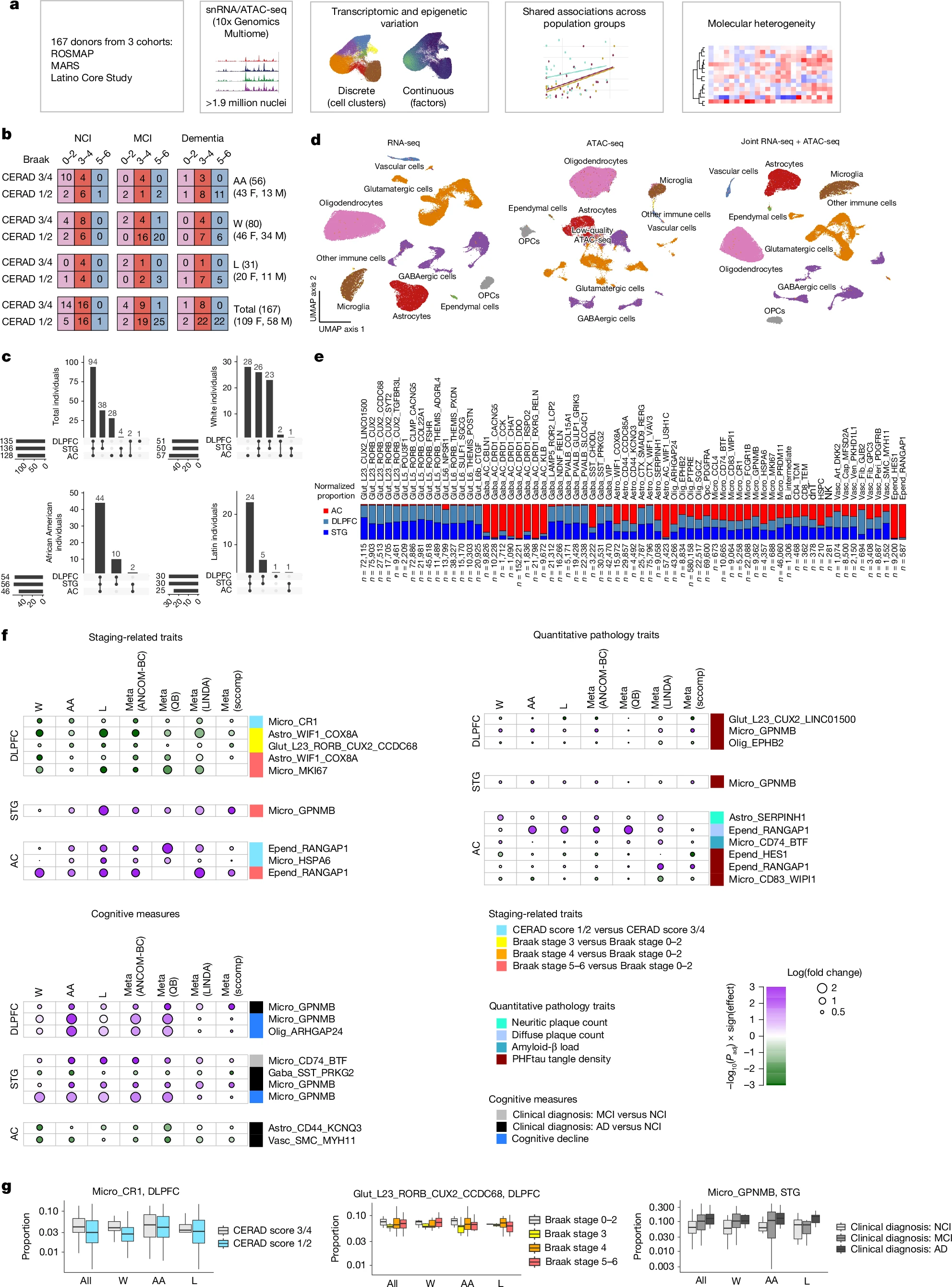
a, Overview of the experimental and analytical workflow. b, Breakdown of pathological stages, cognitive diagnosis and self-reported racial and ethnic groups of individuals in this study. AA, African American (excluding Latin), L, Latin; W, white (excluding Latin). F, female; M, male. c, Upset plot showing the total number of individuals and number of individuals per population group with combinations of brain regions profiled. d, Uniform manifold approximation and projection (UMAP) visualization of nuclei (after quality control) based on snRNA-seq (left), snATAC-seq (middle) and joint snRNA-seq + snATAC-seq (right) data. OPCs, oligodendrocyte precursor cells. e, Relative abundance of each cell-type subcluster across each of the three regions (double-normalized to sum to 1 for each cluster). Astro, astrocytes; dnT, double-negative T cells; Epend, ependymal cells; Gaba, GABAergic neurons; Glut, glutamatergic neurons; HSPC, haematopoietic stem and progenitor cells; Micro, microglia; NK, natural killer cells; Olig, oligodendrocytes; TCM, T central memory; TEM, T effector memory; Vasc, vascular cells. f, P values and effect sizes for all subclusters with statistically significant (meta-analysis FDR-adjusted P < 0.1, consistent directionality and meta-analysis I2 < 50% in at least two out of four compositional analysis methods) associations with pathological and cognitive phenotypes across all three population groups. g, Distributions of specific clusters showing associations with AD phenotypes. Left, the CR1+ myeloid cluster is consistently less abundant in the DLPFC in individuals with CERAD scores 1 and 2 than in those with scores 3 and 4.
The researchers analyzed both cortical and subcortical regions of the brain and identified several cell populations that were consistently associated with Alzheimer’s disease across all three populations. These included specific subgroups of microglia, the brain’s immune cells, astrocytes, which help support neurons, and several types of neurons involved in communication within the brain.
Beyond identifying distinct cell types, the researchers also discovered continuous patterns of gene activity within astrocytes and oligodendrocytes that were strongly associated with Alzheimer’s disease. These molecular programs involved biological processes such as lipid metabolism and neurotransmitter recycling, changes that were not apparent when cells were grouped into traditional categories.
One particularly interesting finding was the identification of six molecular subgroups of individuals with cognitive impairment. These groups were defined by patterns of gene expression rather than by conventional measures of brain pathology, suggesting that Alzheimer’s disease may develop through multiple biological pathways that cannot be detected using standard diagnostic approaches alone.
The results also demonstrate the importance of including diverse populations in biomedical research. Although many disease-associated molecular signatures were shared across ancestry groups, the broader analysis provided a more complete picture of Alzheimer’s disease biology and helped identify common cellular mechanisms that might otherwise have been missed.
As RNA sequencing technologies continue to improve, studies like this are helping researchers understand Alzheimer’s disease at the level of individual cells. Identifying shared and population-specific molecular signatures could improve the development of future biomarkers and therapies while ensuring that discoveries are relevant to more diverse patient populations.
Luquez T, Algoo J, Chiu R, Mares JA, Yadav A, Lam M, Gaur P, Lai X, Lee DI, Paryani F, Batchelor R, Belli I, Henry J, Hoter-Ishay B, Mattison C, Starr L, Lama T, Karaahmet B, Cao W, De Jager PL, Taga M, Barnes LL, Marquez DX, Bennett DA, Zhang Y, Menon V. (2026) Cell-type signatures of Alzheimer’s disease shared across population groups. Nature [Epub ahead of print]. [article]
Related Posts






Alzheimer’s disease affects millions of people worldwide, but most genetic and molecular research has focused on relatively limited populations. Because people have different genetic backgrounds and environmental exposures, scientists have wondered whether the same disease-related cell changes occur across diverse populations or whether important differences have been overlooked.
Researchers at Columbia University Irving Medical Center used single-nucleus RNA sequencing and single-cell chromatin accessibility analysis to investigate Alzheimer’s disease in brain tissue collected from individuals of Latin, African American, and non-Latin white ancestry. Their findings reveal several disease-associated cell types and molecular programs that are shared across all three population groups while also highlighting important differences in how Alzheimer’s disease develops.
Single-nucleus RNA sequencing allows researchers to measure gene activity in individual cells from frozen tissue samples, making it possible to identify the specific cell populations involved in disease. The team paired this approach with ATAC sequencing, which measures how accessible different regions of DNA are for gene regulation. Together, these technologies provide a detailed picture of how brain cells change during Alzheimer’s disease.
Shared associations of discrete clusters with clinical and pathological traits across population groups
a, Overview of the experimental and analytical workflow. b, Breakdown of pathological stages, cognitive diagnosis and self-reported racial and ethnic groups of individuals in this study. AA, African American (excluding Latin), L, Latin; W, white (excluding Latin). F, female; M, male. c, Upset plot showing the total number of individuals and number of individuals per population group with combinations of brain regions profiled. d, Uniform manifold approximation and projection (UMAP) visualization of nuclei (after quality control) based on snRNA-seq (left), snATAC-seq (middle) and joint snRNA-seq + snATAC-seq (right) data. OPCs, oligodendrocyte precursor cells. e, Relative abundance of each cell-type subcluster across each of the three regions (double-normalized to sum to 1 for each cluster). Astro, astrocytes; dnT, double-negative T cells; Epend, ependymal cells; Gaba, GABAergic neurons; Glut, glutamatergic neurons; HSPC, haematopoietic stem and progenitor cells; Micro, microglia; NK, natural killer cells; Olig, oligodendrocytes; TCM, T central memory; TEM, T effector memory; Vasc, vascular cells. f, P values and effect sizes for all subclusters with statistically significant (meta-analysis FDR-adjusted P < 0.1, consistent directionality and meta-analysis I2 < 50% in at least two out of four compositional analysis methods) associations with pathological and cognitive phenotypes across all three population groups. g, Distributions of specific clusters showing associations with AD phenotypes. Left, the CR1+ myeloid cluster is consistently less abundant in the DLPFC in individuals with CERAD scores 1 and 2 than in those with scores 3 and 4.
The researchers analyzed both cortical and subcortical regions of the brain and identified several cell populations that were consistently associated with Alzheimer’s disease across all three populations. These included specific subgroups of microglia, the brain’s immune cells, astrocytes, which help support neurons, and several types of neurons involved in communication within the brain.
Beyond identifying distinct cell types, the researchers also discovered continuous patterns of gene activity within astrocytes and oligodendrocytes that were strongly associated with Alzheimer’s disease. These molecular programs involved biological processes such as lipid metabolism and neurotransmitter recycling, changes that were not apparent when cells were grouped into traditional categories.
One particularly interesting finding was the identification of six molecular subgroups of individuals with cognitive impairment. These groups were defined by patterns of gene expression rather than by conventional measures of brain pathology, suggesting that Alzheimer’s disease may develop through multiple biological pathways that cannot be detected using standard diagnostic approaches alone.
The results also demonstrate the importance of including diverse populations in biomedical research. Although many disease-associated molecular signatures were shared across ancestry groups, the broader analysis provided a more complete picture of Alzheimer’s disease biology and helped identify common cellular mechanisms that might otherwise have been missed.
As RNA sequencing technologies continue to improve, studies like this are helping researchers understand Alzheimer’s disease at the level of individual cells. Identifying shared and population-specific molecular signatures could improve the development of future biomarkers and therapies while ensuring that discoveries are relevant to more diverse patient populations.
Luquez T, Algoo J, Chiu R, Mares JA, Yadav A, Lam M, Gaur P, Lai X, Lee DI, Paryani F, Batchelor R, Belli I, Henry J, Hoter-Ishay B, Mattison C, Starr L, Lama T, Karaahmet B, Cao W, De Jager PL, Taga M, Barnes LL, Marquez DX, Bennett DA, Zhang Y, Menon V. (2026) Cell-type signatures of Alzheimer’s disease shared across population groups. Nature [Epub ahead of print]. [article]
Related Posts
Recent Posts
 Single-cell RNA sequencing links genetic mutations to cell behavior in cancer
 Small RNA sequencing reveals regulatory roles for sdRNAs in acute myeloid leukemia
 Creative Biolabs expands advanced single-cell multi-omics and RNA sequencing solutions to support biomedical discovery
 POND-seq enables non-destructive RNA sequencing in living cells
 Worm’s radical transformation shows metamorphosis can change the functions of cells
 HeartBeat.bio and Cubase Bio Partner to Advance 3D Spatial Transcriptomics for Cardiovascular Drug Discovery
RNA-Seq Products & Services



Stay Connected